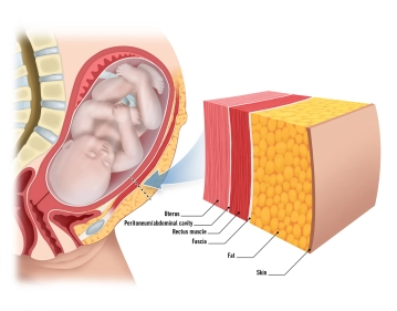
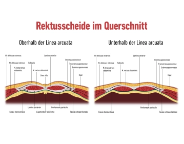
Start your free 3-day trial — no credit card required, full access included

Anatomy - Cesarean section

  1. Skin and subcutaneous tissue

    Skin and subcutaneous tissue

    The skin consists of two main layers:

    1. Epidermis: The outermost layer, composed of keratinized squamous epithelium. It provides protection against physical and chemical influences.
    2. Dermis: Located beneath the epidermis and contains blood vessels, nerves, sebaceous and sweat glands, as well as hair follicles. This layer is responsible for the elasticity and strength of the skin.

    Subcutaneous fat tissue: This layer beneath the dermis serves as a buffer zone and energy reserve. It contains:

    1. Adipocytes: Fat cells that dominate the subcutaneous layer.
    2. Blood vessels and nerves: They run through this layer and can vary individually. Superficial veins and smaller arteries are located here.
  2. Fascia

    Fascia

    Rectus Sheath:

    The rectus sheath is a firm, tendinous structure that envelops the anterior and posterior abdominal muscles. It is formed from the aponeuroses of the abdominal musculature:

    External oblique muscle: The aponeurosis of this muscle forms a significant part of the anterior rectus sheath.

    Internal oblique muscle: This aponeurosis splits above the arcuate line into an anterior and posterior portion, forming both the anterior and posterior layers of the rectus sheath.

    Transversus abdominis muscle: Its aponeurosis reinforces the posterior layer of the rectus sheath above the arcuate line. Below this line, all aponeuroses contribute to the anterior layer.

    The rectus sheath is connected at the linea alba, a vertical tendinous structure extending from the xiphoid process of the sternum to the pubic symphysis.

    Important anatomical structures run within the rectus sheath, such as the superior epigastric artery and vein (above the arcuate line) and the inferior epigastric artery and vein (below the arcuate line). These blood vessels supply the abdominal wall and must be preserved during surgical procedures.

    The rectus sheath provides stability and protection for the underlying rectus abdominis muscle as well as the intra-abdominal contents and is essential for the integrity of the abdominal wall.

    Along the linea alba, a tendinous connection between the rectus sheaths, a surgical approach can be created. This region is relatively avascular, minimizing bleeding.

  3. Muscle tissue

    Muscle tissue

    M. rectus abdominis:

    Origin: The muscle originates from the pubic symphysis and the pubic crest.

    Insertion: Inserts at the costal cartilage of the 5th to 7th ribs and the xiphoid process.

    Function: Primarily responsible for trunk flexion; increases intra-abdominal pressure during abdominal press and stabilizes the upper body.

    Special feature: The muscle is segmented by the tendinous intersections, creating its characteristic appearance. During surgical procedures, it is typically not incised but carefully retracted.

    M. pyramidalis:

    Origin: Originates at the anterior part of the pubic bone and the pubic symphysis.

    Insertion: Inserts into the linea alba, which it tenses.

    Function: Stabilizes the central abdominal wall structure and provides tension in the linea alba.

    Variability: The muscle may be absent, which does not cause functional limitations.

  4. Peritoneum

    Parietal Peritoneum:

    This layer lines the inside of the abdominal wall and forms the outer boundary of the abdominal cavity.

    It is a smooth, serous membrane that provides mechanical protection and facilitates the movement of the underlying organs.

    During a cesarean section, the parietal peritoneum is incised to gain access to the abdominal cavity.

    Visceral Peritoneum:

    It covers the organs in the abdominal cavity, including the uterus (Corpus uteri).

    In the area of the lower uterine segment, it lies particularly close as it envelops the anterior uterine wall.

    Before a hysterotomy, this peritoneum is carefully incised and mobilized.

    The bladder is located immediately in front of the lower uterine segment, separated by the vesicouterine peritoneum. This must be carefully detached from the uterus and mobilized downward during the operation to avoid injury.

  5. The uterus

    The uterus

    The uterus, also known as the womb, is a hollow, muscular organ in the female body that plays an important role during pregnancy and childbirth.

    The uterus consists of:

    • the body (Corpus uteri), 
    • the isthmus uteri
    • the cervix (Cervix uteri) 

    The average length of the uterus is about 7.5 cm, the width 4 cm, and the thickness 2.5 cm. 

    The fundus: uteri represents the upper section of the uterus, located above the entry points of the fallopian tubes. Its lateral walls are connected with the broad ligament (Ligamentum latum uteri). At the lateral edges of the uterus, where the fallopian tubes enter the uterus, the tubal angle is formed. From the tubal angle, the proper ovarian ligament extends to the ovary, while the round ligament of the uterus extends to the inguinal canal.

    Cervix uteri: The cervix extends with its conical lower third into the vagina. It is divided into the supravaginal portion of the cervix (Endocervix) and the vaginal portion of the cervix (Ectocervix or simply Portio). In the upper area of the vagina, the cervix uteri forms the anterior and posterior vaginal fornices (Fornix vaginae) between the Portio and the vaginal wall.

    Portio vaginalis: At the end of the Portio vaginalis is the external os, also known as the Ostium uteri (Orificium externum uteri).

    The Portio vaginalis normally has a reddish color and is covered with stratified, non-keratinized squamous epithelium, similar to the vagina. At the transition to the Ostium uteri, this epithelium changes to the single-layered, columnar epithelium of the cervical canal (Canalis cervicis). Between puberty and menopause, the cylindrical epithelium of the cervical canal can migrate outward onto the Portio (ectropion), leading to more intense redness. With a colposcopy, precancerous lesions and cancer can be detected in this area.

    Endometrium: This inner mucosal layer undergoes significant proliferation and differentiation to provide an optimal environment for the implantation and nourishment of the embryo. During pregnancy, it forms the functional basis for the placenta, which ensures gas exchange and nutrient supply.

    Myometrium: The middle muscular layer consists of smooth muscle and is organized into three layers (longitudinal, circular, and oblique). During pregnancy, the myometrium increases in both mass and length to accommodate the growing fetus. At the end of pregnancy, the uterus weighs about 1,000 grams (compared to 50-70 grams in a non-pregnant state) and has a volume of about 5 liters.

    In the lower uterine segment, the myometrium becomes thinner and more elastic, facilitating surgical access during a cesarean section.

  6. Placenta

    Placenta

    The placenta is a disc-shaped organ that forms during pregnancy and serves as an essential connection between the mother and fetus. It is approximately 2-3 cm thick, has a diameter of 15-20 cm, and weighs about 500-600 grams at birth.

    Maternal side (basal plate): This side is adjacent to the uterine wall and consists of the decidua basalis. It contains spiral arteries that supply the placenta with blood, facilitating the exchange of oxygen and nutrients.

    Fetal side (chorionic plate): The fetal side is formed by the chorionic plate and is covered with the amniotic membrane. It contains chorionic villi that extend into the intervillous spaces, where the exchange between maternal and fetal blood occurs.

    Maternal blood flows through spiral arteries into the intervillous spaces, where gases and nutrients diffuse. Fetal blood is enriched with oxygen via the umbilical vein (vena umbilicalis) and transported back to the placenta through two umbilical arteries (arteriae umbilicales).

  7. Adnexa

    Adnexa

    Ovary (Ovarium)

    The ovary (Ovarium) is located laterally in the pelvic region and runs in a vertical orientation. It has an inner surface facing the pelvic interior (Facies medialis/intestinalis) and an outer surface directed towards the lateral pelvic wall (Facies lateralis). The rounded free edge is referred to as the Margo liber, and at the anterior edge, the Margo mesovaricus, the mesovarium is attached, which is a structure of the broad ligament of the uterus (Lig. latum uteri).

    There are two poles: 

    • Uterine extremity
    • Tubal extremity

    The hilum of the ovary, where vessels and nerves enter, is located at the attachment of the connective tissue of the ovary, known as the mesovarium, at the Margo mesovaricus.

    The ovary is connected to the pelvic wall at the tubal extremity by the suspensory ligament of the ovary. The ovarian vessels (Vasa ovarica), lymphatic vessels, and nerves run along this suspensory ligament.

    The proper ovarian ligament lies between the ovary (uterine extremity). This is located directly behind the tubal angle. The artery R. ovaricus of the uterine artery runs here. 

    The ovary is supplied by: 

    • Ovarian artery
    • Ovarian branch from the uterine artery.

    These arteries form anastomoses at the Margo mesovaricus and supply the ovary with blood. 

    Veins transport the venous blood via the right ovarian vein to the inferior vena cava and via the left ovarian vein to the left renal vein. Part of the blood flows through the uterine plexus to the internal iliac vein.

    The ovary is located in the abdominal cavity (intraperitoneal) in the ovarian fossa. Here, in the retroperitoneum, run the obturator nerve, obturator vessels, and external iliac vessels. Medially, the umbilical artery and uterine artery also run.


    The Fallopian Tube (Tuba uterina, Salpinx)

    The fallopian tube, also known as the Tuba uterina, is approximately 10-15 centimeters long and 2-5 millimeters thick. It runs intraperitoneally along the upper free edge of the broad ligament of the uterus (Ligamentum latum uteri), from the corner of the uterus to the ovary. In the mesosalpinx, the blood vessels and nerves to the fallopian tube run, additionally anchoring the fallopian tube to the broad ligament.

    The fallopian tube itself can be divided into different sections:

    • Infundibulum of the fallopian tube (+ abdominal ostium of the fallopian tube)
    • Fimbriae of the fallopian tube, 
    • Ampulla of the fallopian tube
    • Isthmus of the fallopian tube
    • Uterine part. 

    The blood supply of the fallopian tube is provided by small branches (Rr. tubarii) from the ovarian and uterine arteries, which run in the mesosalpinx and anastomose with each other. Veins accompany the arteries and drain into the ovarian vein and the uterine/uterovaginal plexus. 

  8. Bladder and Ureter

    Located directly in front of the lower uterine segment, the bladder is separated from the uterus by the vesicouterine peritoneum. During a cesarean section, it is carefully moved downward to avoid injury. This is particularly important in complications such as a low-lying placenta or adhesions resulting from previous surgeries.

    Consists of a robust muscle layer, the detrusor muscle, and is lined with mucosa. The upper part of the bladder is covered with peritoneum, while the lower part is extraperitoneal.

    The ureter runs retroperitoneally from the renal pelvis through the small pelvis to the bladder. In the area of the uterus, it runs close to the uterine artery, about 1-2 cm lateral to the transition to the cervix. The ureter can be injured by deep surgical procedures or in the presence of adhesions.