Start your free 3-day trial — no credit card required, full access included

Anatomy - Right Hemihepatectomy, Robotically Assisted

  1. Functional liver anatomy

    Functional liver anatomy

    The liver is macroscopically divided into a larger right lobe and a smaller left lobe (volume ratio approximately 80:20) by the ligamentum falciforme, the insertion of the ligamentum teres hepatis on the diaphragmatic surface, and the fissura sagittalis on the visceral surface. However, this morphological division does not correspond to the liver’s functional structure.

    The functional segmentation of the liver is determined by the branching patterns of the portal triad structures: the portal vein (vena portae), the hepatic artery (arteria hepatica), and the bile duct (ductus choledochus). These structures branch in a consistent pattern both at the porta hepatis and within the parenchyma. Each liver segment is completely independent in terms of blood supply and bile drainage, allowing any segment to be surgically removed without compromising the functionality of the remaining liver.

    The concept of functional anatomy refers to this subdivision of the liver into hemodynamically independent parenchymal units, knowledge of which is critical for planning and executing liver resection procedures.

  2. Portal vein and hepatic veins

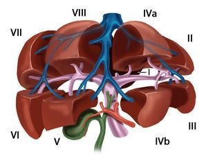
    The functional division of the liver is based on the branching of the portal vein into independent subunits called segments.

    Portal Vein Anatomy:

    • The portal vein typically bifurcates at the liver hilum into a right and left main branch
    • The boundary between the territories of these branches is the Cava-Gallbladder line (Cantlie’s line)
    • The right branch divides further into an anteromedial trunk (supplying segments V/VIII) and a posterolateral trunk (supplying segments VI/VII)
    • The left branch courses transversely to the left and then anteriorly as the pars umbilicalis, terminating at the insertion of the ligamentum teres hepatis in the Recessus rex. It provides branches to:
      • Segments II and III (left lateral segments)
      • Segments IVa and IVb (left medial segments)

    Unique Case of the Caudate Lobe:

    The Lobus caudatus (Segment I) is unique as it receives significant inflow from both the right and left main portal branches.

    Segmentation According to Couinaud:

    The liver is divided into eight portal venous segments, numbered clockwise starting from the caudate lobe:

    • Segment I: Caudate lobe
    • Segments I/II/III: Left lateral lobe
    • Segment IV: Left paramedian sector (Lobus quadratus)
    • Segments I/II/III/IV: Left liver lobe
    • Segments V/VIII: Right paramedian sector
    • Segments VI/VII: Right lateral sector
    • Segments V/VI/VII/VIII: Right liver lobe

    Hepatic Veins:

    The liver is traversed by three main hepatic veins — right, middle, and left hepatic veins — which divide it into four hepatic sectors:

    • The left hepatic vein drains the left lateral lobe and usually joins the middle hepatic vein near the vena cava
    • The middle hepatic vein follows Cantlie’s line and separates the left and right paramedian sectors
    • The right hepatic vein runs between the posterolateral and anteromedial segment

    The Lobus caudatus has independent venous drainage through multiple small veins (Spieghel’s veins) that directly empty into the vena cava.

    Portal and Venous Hilum:

    • The portals of segments II, III, and IV are extrahepatic and can be dissected relatively easily at the anterior portion of the left umbilical fissure
    • The portals of right-sided liver segments are intrahepatic, with Segment VI occasionally presenting exceptions
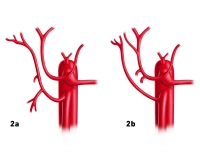
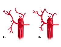
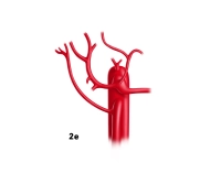
    Variations:

    Portal Venous System:

    • Anomalies of portal vein bifurcation almost exclusively affect the right main portal branch:
      • Portal vein trifurcation: The right main branch is absent, replaced by two branches supplying segments V/VIII and VI/VII
    • In rare cases, one right branch may arise from the left portal vein
    • Variants in the left portal system usually involve segmentation:
      • Multiple small branches instead of two distinct branches for IVa/IVb
      • Occasionally, an intermediate branch exists between segments II and III
    40_LAV_01_a_b_Pfortader_Lebervenen

    Figures 1a and 1b

    • PV = Portal vein
    • LPV = Left portal vein
    • RA = Right anterior portal vein branch
    • RP = Right posterior portal vein branch

    Hepatic Veins:

    • Variations in hepatic veins are more common than in the portal vein system
    • Deviations from Couinaud’s hepatic sectors primarily affect the territories of the right and middle hepatic veins

    This detailed knowledge of functional and variant anatomy is essential for precise planning and execution of liver resections.

  3. Hepatic arteries

    The common hepatic artery arises from the celiac trunk, though in rare cases, it may originate directly from the aorta or the superior mesenteric artery. After giving off the gastroduodenal artery, it continues as the proper hepatic artery, which bifurcates at the liver hilum into the right and left hepatic arteries.

    Extrahepatic branches, such as the artery supplying Segment IV, often arise just before the umbilical fissure from the left hepatic artery.

    Variations:

    • Deviations from the normal arterial pattern occur in 30 % of cases
    • Common anatomical variations include:
      • A replaced or accessory hepatic artery originating from the superior mesenteric artery or the left gastric artery
    • Both variations may occur simultaneously
    40_LAV_02_a_b_Leberarterie
    40_LAV_02_c_d_Leberarterie
    40_LAV_02_e_Leberarterie
  4. Biliary System

    Left Hepatic Duct (Ductus hepaticus sinister)

    • The extrahepatic portion of the left hepatic duct measures approximately 3 – 5 cm and forms in the umbilical fissure from the union of ducts draining Segments II and III
    • The bile duct from Segment IV typically joins proximally to this union
    40_LAV_03_a_Gallenwege

    Right Hepatic Duct (Ductus hepaticus dexter)

    • The right hepatic duct is short, approximately 1 cm, and collects bile through anterior and posterior branches from Segments V, VI, VII, and VIII
    • In some cases, the right hepatic duct is absent, and anterior/posterior branches drain directly into the common hepatic duct
    40_LAV_03_b_Gallenwege

    Variations:

    Left Hepatic Duct

    • The most frequent and clinically significant variation (~ 25 %) is a common union of the bile duct from Segment IV with those of Segments II and III
    • This variation is critical during left lateral resections, living donor liver transplantation, and liver splitting, as it can impair bile drainage from Segment IV

    Right Hepatic Duct

    • It may be very short or absent, with anterior and posterior pedicles draining directly into the common hepatic duct. This eliminates the typical hepatic duct bifurcation

    Figures 3b to 3g

    • rp = Right posterior bile duct
    • ra = Right anterior bile duct
    •  dhs = Left hepatic duct

    Biliary Confluence Variations

    • Posterior branch drains directly into the cystic duct, common hepatic duct, or common bile duct (choledochus)
    • Anterior or more commonly posterior branches of the right hepatic duct drain into the left hepatic duct
    40_LAV_03_c_Gallenwege
    40_LAV_03_d_Gallenwege

    Clinical Relevance:

    • During a left hemihepatectomy, accidental transection of such variant ducts can result in severe bile leakage and segmental cholestasis in the right liver lobe
    40_LAV_03_e_Gallenwege
    40_LAV_03_f_Gallenwege
    40_LAV_03_g_Gallenwege

    Understanding these arterial and biliary anatomical variations is essential to avoid complications during hepatic resections, liver transplantation, and biliary interventions.

  5. Regional Lymph Nodes

    The liver has two main lymphatic drainage pathways:

    Primary Pathway (90 %)

    • Lymph from the liver flows to the lymph nodes at the porta hepatis
    • From there, it drains via the celiac lymph nodes (Nodi lymphatici coeliaci) into the intestinal trunk and eventually to the thoracic duct

     Secondary Pathway (10 %)

    • This pathway serves the surface of the diaphragmatic face and the area nuda of the liver
    • Lymph passes through the diaphragm into the superior phrenic lymph nodes and then via mediastinal lymphatics into the right venous angle