Start your free 3-day trial — no credit card required, full access included

Anatomy - Myotomy and Fundoplication according to Dor, robotically assisted

  1. Surgical Anatomy of the Esophagus and Stomach

    Surgical Anatomy of the Esophagus and Stomach 1
    Surgical Anatomy of the Esophagus and Stomach 2

    Esophagus

    General Characteristics

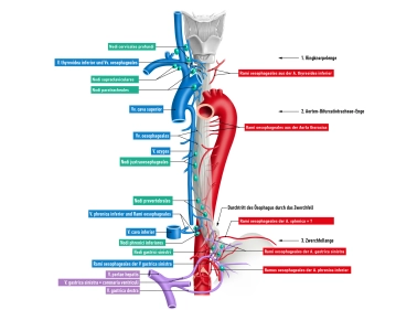
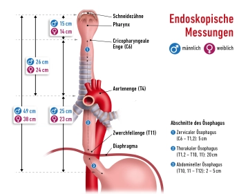
    • Muscular hollow organ, approx. 25–30 cm long
    • Connects pharynx (C6) with the stomach (Th11)
    • Three constrictions:
      1. Upper constrictioncricoid constriction (transition pharynx/esophagus, at level C6)
      2. Middle constrictionaortic constriction (crossing with aortic arch + left main bronchus)
      3. Lower constrictiondiaphragmatic constriction (esophageal hiatus, Th10)

    Topographical Sections

    Cervical Esophagus (C6–Th1)

    • Behind the trachea
    • Accompanying structure: recurrent laryngeal nerve
    • Access: transcervical, ventral-lateral

    Thoracic Esophagus (Th1–Th10)

    • Upper mediastinum: behind trachea, in front of spine
    • Middle mediastinum: behind heart and pericardium
    • Crossing by aortic arch, azygos vein, left main bronchus
    • Access: right-thoracic (better overview)

    Abdominal Esophagus (short, 1-3 cm)

    • Passes through esophageal hiatus (Th10) into the abdomen
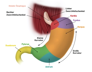
    • Empties into the cardia region of the stomach
    • Accompanying structures: anterior vagal trunk (left), posterior (right)


    Stomach

    General & Location

    • Muscular hollow organ between esophagus and duodenum
    • Located in the left/middle upper abdomen, directly under the diaphragm
    • Can vary greatly in size and shape depending on age, filling state, and body position
    • Great interindividual differences in terms of location, size, and shape

    Size & Filling Capacity

    • Average length: 25–30 cm (with moderate filling)
    • Storage capacity: approx. 1.5 liters, in extreme cases up to 2.5 liters

    Stomach Layers and Attachment

    • Located intraperitoneally; mostly covered with serosa (except dorsal cardia)
    • Embryonic mesogastria rotate into a frontal position:
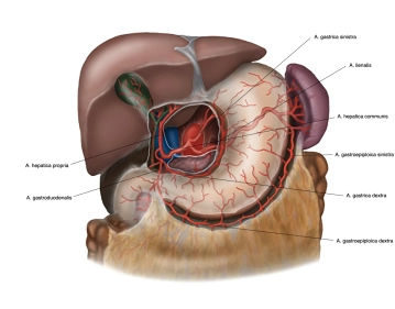
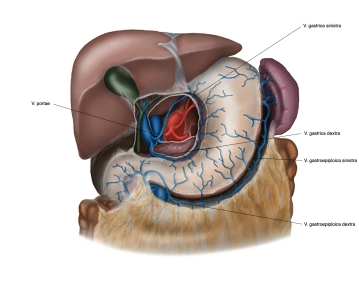
      • Lesser omentum: from the lesser curvature to the hepatic portal
      • Greater omentum: from the greater curvature to the transverse colon, spleen, and diaphragm
    • Attachment and stabilization by ligaments that, among other things, extend to the liver and spleen

    Sections of the Stomach

    • Cardia (stomach entrance, upper gastric orifice, ostium cardiacum)
      • Area of 1–2 cm in which the esophagus empties into the stomach
      • Distinct transition from esophageal mucosa to gastric mucosa (clearly visible on endoscopy)
    • Gastric Fundus (stomach fundus)
      • Located above the stomach entrance, bulges upward
      • Also referred to as gastric dome or fornix gastricus
      • Typically filled with air; in upright position the highest point, recognizable as “gastric bubble” on X-ray
      • Delimited from the stomach entrance by the cardiac notch
    • Gastric Corpus (stomach body)
      • Main part of the stomach
      • Characterized by deep longitudinal mucosal folds (plicae gastricae) that run from the stomach entrance to the pylorus (“gastric canal”)
    • Pylorus (pyloric part, pyloric sphincter)
      • Beginning with the expanded pyloric antrum, followed by pyloric canal (canalis pyloricus)
      • Ends with the actual pyloric sphincter (pylorus), where the pyloric sphincter muscle (M. sphincter pylori) is located
      • Closes the stomach outlet (ostium pyloricum) and regulates the passage of chyme into the duodenum

    Formal Boundaries and Further Anatomical Features

    • Greater curvature (convex side)
    • Lesser curvature  (concave side)
    • Anterior wall: Paries anterior; Posterior wall: Paries posterior
    • Greater omentum originates from the greater curvature; Lesser omentum spans between the left hepatic lobe and the lesser curvature
  2. Layers and Structure of the Esophagus and Stomach Wall/Microscopy

    Layer structure of the esophagus (from inside to outside)

    1. Tunica mucosa (Mucosa)

    • Lamina epithelialis mucosae: multilayered, non-keratinized squamous epithelium – mechanically protects against food and gastric acid, with a clear Z-line to the gastric epithelium 
    • Lamina propria mucosae: loose connective tissue with small vessels, lymphoid tissue, and nerves 
    • Lamina muscularis mucosae: thin smooth muscle layer – contracts independently of the muscularis propria 

    2. Tela submucosa

    • Connective tissue layer with Glandulae oesophageales (mucous glands), which produce mucus for the swallowing process 
    • Meissner plexus: enteric nerve plexus for controlling mucosal activity
    • Longitudinal folds in the tissue allow expansion during swallowing; in cross-section, the lumen appears star-shaped 

    3. Tunica muscularis (Muscle wall)

    • Consists of two layers:
      • Stratum circulare (circular muscle)
      • Stratum longitudinale (longitudinal muscle)
    • Muscle distribution:
      • upper third: striated muscle
      • middle third: mixed muscle
      • lower third: exclusively smooth muscle
    • Auerbach plexus (myenteric plexus) between the muscle layers: coordination of peristalsis 

    4. Tunica adventitia / Tunica serosa

    • Pars thoracica: Adventitia – loosely connecting, collagenous connective tissue, fixes the esophagus in its environment 
    • Pars abdominalis (after passage through the diaphragm): covered by serosa – intraperitoneal segment with single-layered squamous epithelium 


    Layers of the stomach wall from inside to outside
    Under the microscope, the stomach wall shows a characteristic layer structure from inside to outside:

    1. The Tunica mucosa is the mucosal layer that lines the stomach from the inside.  The gastric mucosa is divided into three sublayers: The lamina epithelialis mucosae forms a tough neutral mucus that protects the gastric mucosa from mechanical, thermal, and enzymatic damage. Below that follows the lamina propria mucosae as a sliding layer, in which the gastric glands (Glandulae gastricae) are embedded. Finally, there is a narrow lamina muscularis mucosae, a thin first muscle layer that can change the relief of the mucosa.
    2. The Tela submucosa follows in the view from inside to outside of the gastric mucosa. It represents a loose sliding layer consisting of connective tissue. In the tela submucosa, there is a dense network of blood and lymph vessels, as well as a nerve fiber plexus, the submucosal plexus (Meissner plexus), which controls gastric secretion. This plexus works independently of the central nervous system (CNS) and is influenced by the autonomic nervous system.
    3. This is followed by a strong Tunica muscularis, which is divided into three sublayers with fibers running in different directions: an inner layer of small obliquely running muscle fibers (Fibrae obliquae), then a circular muscle layer (Stratum circulare), and outermost an outer longitudinal muscle layer (Stratum longitudinale). This musculature is responsible for the peristalsis of the stomach, which is responsible for the constant mixing of the chyme with the gastric juice. Between the circular and longitudinal muscle layers runs a nerve fiber plexus, the myenteric plexus (Auerbach plexus), which controls the function of the musculature. Just like the submucosal plexus, this plexus works largely autonomously but is influenced by the autonomic nervous system.
    4. This is followed by another connective tissue sliding layer (Tela subserosa).
    5. The conclusion is formed by the Tunica serosa. The serosa is also divided into several layers. The tela subserosa is followed by the Lamina propria serosae. In this, the blood and lymph vessels as well as nerves run. In addition, there are cells of the immune defense in the lamina propria, which are referred to as milky spots (Macula lactea). The Lamina epithelialis serosae is finally directed towards the body cavity and consists of a single-layered squamous epithelium, the serosal epithelium.This layer is shiny, transparent, and ensures good slidability of the stomach relative to the adjacent organs through a thin fluid film. 

    Gastric glands
    The gastric glands (Glandulae gastricae) are located in the lamina propria mucosae and can be found in the fundus and corpus of the stomach. Up to 100 glands are found per 1mm2 of the mucosal surface. Various cells are located in the wall of the glandular tube:

    • Mucus cells: They produce the same neutral mucus as the epithelial cells.
    • Accessory cells: These cells are located quite superficially in the gland and secrete alkaline mucus, i.e., the pH value is high due to the bicarbonate ions (HCO3- ions) contained therein. This property is important for controlling and, if necessary, regulating the pH value of the stomach. The mucus coats the gastric mucosa and thus protects against self-digestion by the aggressive hydrochloric acid (HCl) and enzymes as self-digesting proteins. This cell type is found in abundance in the cardia and fundus of the stomach.
    • Chief cells: These cells produce the inactive precursor enzyme pepsinogen, which is converted into the active enzyme pepsin by hydrochloric acid (HCl) after release and is responsible for the digestion of food proteins. Since the enzyme only comes into contact with the hydrochloric acid at the surface of the gland, self-digestion of the glands by the enzyme is prevented. This cell form is mainly found in the corpus of the stomach.
    • Parietal cells: These cells, which are found in greater numbers in the gastric corpus, produce abundant hydrogen ions (H+ ions), which are needed for the formation of hydrochloric acid (HCl). The hydrochloric acid has a very low pH value of 0.9-1.5. In addition, the parietal cells form the so-called intrinsic factor. This substance forms a complex in the intestine with vitamin B12 from food, which can then pass through the small intestinal wall. This vitamin is of particular importance in erythropoiesis (removal of the stomach can lead to anemia).
    • G-cells: These cells, which are preferably located in the antrum of the stomach, produce gastrin to increase HCl formation in the parietal cells.
  3. Functions of the Esophagus and Stomach

    Esophagus Function

    • Transport of Food and Liquid
      Peristaltic waves of the longitudinal and circular muscles transport the bolus from the hypopharynx to the stomach. In this process, the swallowing act is initially voluntary, then reflexive.
    • Protection against Reflux
      Functional closure by the lower esophageal sphincter (UES = upper, LES = lower sphincter) as well as the crura of the diaphragm.
    • Controlled Passage through the Diaphragm
      Coordination of swallowing act and respiratory movement.

    Stomach Function

    • The stomach serves as a reservoir for the ingested food. Its task is to store the food and to mix it. 
    • In the stomach, the acidic gastric juice (mucus and HCl) and enzymes are produced, which pre-digest some components of the food. Subsequently, the chyme is passed portionwise through the pylorus into the duodenum. 
    • It can store the food for hours and thus ensures that we can cover our daily nutritional needs with a few larger meals. 
  4. Arterial Supply

    Arterial Supply 1
    517_AN_02_Oesophagus.jpg

    Arterial Vascular Supply Esophagus

    The blood supply of the esophagus occurs segmentally. It receives arterial blood from three main sections, corresponding to its topographic division:

    Section

    Main Arteries

    Origin

    Cervical

    Esophageal branches from the inferior thyroid artery

    Thyrocervical trunk (subclavian artery)

    Thoracic

    Esophageal branches from the thoracic aorta

    + small branches from the bronchial arteries

    Direct aortic branches, bronchial arteries

    Abdominal

    Esophageal branches from the left gastric artery (main supply) 

    + possibly from the left inferior phrenic artery

    Celiac trunk, inferior phrenic artery


    The arterial supply of the stomach occurs via several blood vessels, all of which originate from the unpaired celiac trunk. These run along the gastric curvatures as vascular arcades to supply the organ and form numerous anastomoses among themselves:

    • Right gastric artery from the proper hepatic artery to the lower part of the lesser curvature,
    • Left gastric artery to the upper part of the lesser curvature,
    • Short gastric arteries from the splenic artery to the fundus,
    • Right gastroepiploic (-omental) artery from the gastroduodenal artery to the lower (right) part of the greater curvature,
    • Left gastroepiploic (-omental) artery from the splenic artery to the left side of the greater curvature,
    • Posterior gastric artery from the splenic artery to the posterior wall.

    Through this, the stomach is supplied by 2 vascular arcades between the left and right gastric arteries on the lesser curvature, as well as the left and right gastroepiploic arteries on the greater curvature.

  5. Venous and Neural Supply

    517_Anatomie_venen besch.jpg
    517_AN_02_Oesophagus.jpg

    Venous Drainage of the Esophagus

    The venous drainage of the esophagus occurs segmentally in three directions and forms an important portocaval anastomosis through its connections between the portal and caval venous systems.

    Note: This portocaval anastomosis is clinically particularly significant in portal hypertension.

    • Cervical: V. thyroidea inferior → V. brachiocephalica
    • Thoracic: Vv. oesophageae → V. azygos / hemiazygos
    • Abdominal: V. gastrica sinistra → V. portae
      ➡ Anastomoses between V. azygos and V. portae = portocaval connection (esophageal varices in portal hypertension).


    Neural Supply of the Esophagus

    The neural supply of the esophagus is divided into somatomotor/sensory components for the upper (striated) muscle portion and visceromotor/sensory components for the smooth muscle portion.

    • Motor Function
      • Upper Third (Striated Musculature)
        • Somatomotor: N. vagus (Rr. pharyngei, R. recurrens) → voluntary initiation of swallowing
      • Middle and Lower Third (Smooth Musculature)
        • Visceromotor: N. vagus via the Plexus oesophageus → controlled by parasympathetic reflexes
    • Parasympathetic Innervation
      • Source: Nn. vagi (right and left vagus)
      • Course: Form the Plexus oesophageus in the thorax → pass as Trunci vagales (anterior/posterior) through the diaphragm to the stomach
      • Function: Peristalsis, secretion, sphincter tone
    • Sympathetic Innervation
      • Source: Truncus sympathicus (segments Th1–Th6) → Nn. splanchnici major/minor → Plexus oesophageus
      • Function: Inhibition of peristalsis, increase in sphincter tone
    • Sensory Innervation
      • Somatosensory (pain, touch in the upper third): N. vagus, N. laryngeus recurrens
      • Viscerosensory (distension, chemical stimuli in the lower third): Vagus and sympathetic pathways → posterior horns of the spinal cord
    • Surgical Relevance:
      • Injury to the N. laryngeus recurrens (e.g., during cervical esophageal mobilization) → hoarseness, risk of aspiration
      • Denervation of the lower esophagus in fundoplication or esophagectomy can lead to motility disorders


    Venous Supply of the Stomach

     Parallel to the arterial supply, the 4 major veins of the stomach run along the two curvatures. Overall, they form collecting veins (V. gastrica sinistra and dextra directly into the V. portae hepatis, V. gastroomentalis sinistra and Vv. gastricae breves to the V. splenica, and V. gastroomentalis dextra to the V. mesenterica sup.), which ultimately all drain into the portal vein.


    Neural Supply of the Stomach 

    The neural supply of the stomach is predominantly under the autonomic nervous system. There are also sensory fibers: The sympathetic system supplies the musculature of the pylorus, the parasympathetic system (N. vagus X) supplies the remaining stomach musculature and the glands of the stomach. The N. vagus runs right and left parallel to the esophagus, passes through the diaphragm via the esophageal hiatus and reaches the anterior surface of the stomach on the left side (Tr. vagalis anterior), and the posterior surface on the right side (Tr. vagalis posterior). Sensory fibers from the stomach, however, run afferently via the N. splanchnicus major to the thoracic spinal ganglia.

Lymphatic Drainage Pathways

Lymphatic Drainage Pathways of the EsophagusCervical → paratracheal, deep cervical LNThoraci

Activate now and continue learning straight away.

Single Access

Activation of this course for 3 days.

US$9.50  inclusive VAT

Most popular offer

webop - Savings Flex

Combine our learning modules flexibly and save up to 50%.

from US$4.40 / module

US$52.91/ yearly payment

price overview

robotics

Unlock all courses in this module.

US$8.81 / month

US$105.80 / yearly payment

to top