Start your free 3-day trial — no credit card required, full access included

Perioperative management - Chimney Technique for Juxtarenal Aortic Aneurysm (Ch-EVAR)

  1. Indications

    Approximately 80% of abdominal aortic aneurysms are located infrarenally. However, in up to 20%, there is no sufficient aneurysm neck (juxtarenal aneurysm), and the AAA can also begin at or above the renal arteries (suprarenal aneurysm). The missing or morphologically inadequate infrarenal anchoring zone („hostile neck“) excludes a conventional endovascular aneurysm exclusion.

    Fenestrated prostheses or the chimney technique (Ch-EVAR = „chimney technique with endovascular aneurysm repair“) allow these pathologies to be treated endovascularly. The chimney technique creates a proximalization of the landing zone to ensure the necessary anchoring for the abdominal stent graft. This is achieved by implanting a usually covered stent (chimney graft) into the affected renovisceral vessel of the aorta, parallel and outside the aortic endoprosthesis.

    The indications for the treatment of pararenal pathologies using the chimney technique include:

    • large rupture-prone aneurysms (> 6 cm diameter) or penetrating aortic ulcers without thrombus formation
    • abdominal aneurysms with „hostile neck“ and pronounced angulation/calcification of the iliac arteries and/or the aneurysm neck
    • symptomatic, ruptured pathologies
    • Type Ia endoleak after previous EVAR
    • Presence of relevant, accessory or ectopic renal arteries with a diameter of more than 4 mm
    • paraanastomotic aneurysms after previous open aneurysm treatment

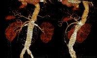
    In the video example, it is a symptomatic abdominal aortic aneurysm with 9.5 cm diameter at the level of the renal arteries (juxtarenal), accompanying CAD, s/p pacemaker implantation right prepectoral:
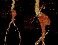
    PM 321-1

    Postoperative:

    PM 321-2
  2. Contraindications

    Because in interventional procedures, due to unexpected technical problems, a change in procedure to an open operation may become necessary, preoperative risk assessment and the definition of contraindications are important:

    • Heart failure (ejection fraction < 25-30 %)
    • COPD (FEV1 < 0,8)
    • non-correctable severe CAD
    • incurable cancer

    Relative contraindication:

    • compensated renal insufficiency (due to intraoperative contrast medium load)

    If in borderline cases the operation is to be performed despite contraindications, this requires special agreement with the patient or with his relatives.    

  3. Preoperative Diagnostics

    Thorough vascular surgical examination:

    • Pulse status
    • Doppler of the legs (frequent coincidence of popliteal aneurysms)
    • if applicable, walking distance test in case of concurrent PAD and poorly palpable foot pulses
    • Abdominal ultrasound
    • Color duplex of the carotids (frequent coincidence of internal carotid stenoses)
    • Echocardiogram
    • Exercise ECG
    • Laboratory investigations (electrolytes, coagulation, renal values, blood count, blood lipids)
    • Chest X-ray
    • if applicable, in the presence of concurrent PAD or critical visceral artery morphology, intra-arterial DSA of the abdominal aorta and pelvic and leg vessels
    • Angio-CT of the thorax, abdomen, and pelvis to assess the vessels of the upper extremities and the descending aorta
  4. Special Preparation

    • General OR Preparations
    • Determine blood group, if necessary provide blood products
    • Duplex sonography of the access vessels (A. brachialis, A. axillaris and if necessary A. subclavia on the access side)
    • Fasting limit 6 hours
    • prophylactic antibiosis is usually performed and is recommended by the German Society for Vascular Surgery (single-shot Cefuroxime 30 minutes before skin incision), however, the benefit is currently being discussed due to the resistance problem (RKI)   
  5. Informed Consent

    General Surgical Risks

    • Severe bleeding, blood transfusions, transmission of hepatitis/HIV through blood products
    • Allergy/intolerance
    • Wound infection
    • Thrombosis/embolism
    • Skin, vascular, nerve damage e.g. due to positioning
    • Keloids (if open access)

    Specific Procedure Risks

    • Vascular injuries e.g. during prosthesis placement: surgical hemostasis, blood transfusions, in case of severe bleeding or aortic tear immediate conversion to open surgery
    • Pseudoaneurysm of the punctured vessels, AV fistula, seroma
    • Aortoenteric fistula: Surgical intervention, possibly with removal of the vascular prosthesis
    • Inadequate anchoring or leakage of the prosthesis: Corrective procedures, possibly open
    • Infection of the vascular prosthesis: possible after days, months or years; Endocarditis,  sepsis, leg ischemia, amputation; surgical removal of the vascular prosthesis
    • Stenosis and occlusion of the inserted chimney stents
    • Damage to adjacent organs; Intestinal ischemia -> Resection, artificial anus placement
    • Nerve lesions -> Sensory disturbances, pain, paralysis of the abdominal wall, thigh muscles
    • Lymph fistula
    • Lymphedema of the legs; temporary, permanent; compression stockings, lymphatic drainage
    • Postoperative bleeding
    • Impotence    
    • Compromising renal function through intraoperative angiography, permanent dialysis

    Risks due to Perfusion Disorders

    • Thrombosis/Embolism: Pulmonary embolism, stroke, myocardial infarction; Prophylaxis Heparin-> HIT II risk
    • Legs: Thrombosis of the vascular prosthesis and possibly adjacent vascular segments, possibly leg ischemia, amputation (e.g. toes)
    • Kidneys: due to thrombosis or placement of the vascular prosthesis; Despite immediate surgical correction possibly permanent kidney damage -> Dialysis
    • Intestine: Perfusion disorders due to thrombosis/embolism and possibly placement of the vascular prosthesis; Vascular surgical intervention required, possibly intestinal resection, permanent artificial anus
    • Spinal cord: Depending on the size of the aneurysm perfusion disorders due to placement of the vascular prosthesis; Temporary sensory disturbances/paresis, possibly also permanent paraplegia in case of low-originating A. radicularis magna
    • Gluteal muscles: due to overstenting of the pelvic arteries, especially bilaterally; Claudication symptoms when walking, possibly gluteal necrosis formation
    • Liver: Functional disorder due to hypoperfusion, especially in pre-existing liver diseases
  6. Anesthesia

    ITN

  7. Positioning

    Positioning
    • Supine position
    • the access arm* is adducted, the opposite side is abducted

    * Access arm is usually left, in the film example however right due to status post pacemaker implantation left!

    For the purpose of intraoperative fluoroscopy, the OR table top should be made of carbon. The carbon table should be free on both sides for possible movements of the C-arm, therefore better to test all swivel maneuvers of the C-arm beforehand.

  8. OR Setup

    OR Setup

    Due to the pacemaker located on the left, in the video example, the surgeon and assistant stand on the right, the OR specialist on the left at the level of the patient's foot end. The C-arm and monitors are located opposite the surgeon/assistant.

  9. Special Instrumentation and Holding Systems

    Special Instrumentation and Holding Systems
    Mögliche Prothesenmodule
    • long 7F- or 8F-sheaths, 20F-sheath
    • Guidewires (Terumo® wire 0.035, Rosen wires, 2 stiff wires such as Back-up-Meier® or Amplatz®)
    • Guiding catheters depending on configuration (Pigtail, Vertebral catheters etc.)
    • modular endoluminal aortic bifurcation prosthesis (measure exactly before surgery), covered stents for the renal arteries depending on diameter and length measured before surgery (best in 2-3 different lengths and diameters)
    • Dilation balloon (6-9 mm with long shaft)
    • Aortic stent (depending on length and diameter of the aorta and the aneurysm if additional central fixation should become necessary)
    • high-performance DSA system, possibly hybrid OR
    • Contrast medium high-pressure pump, possibly CO2 injector
    • for conversion in emergency large vessel sieve
  10. Postoperative Treatment

    Postoperative Analgesia

    Follow the link here to PROSPECT (Procedures Specific Postoperative Pain Management) and to the current guideline Treatment of acute perioperative and posttraumatic pain.

    Medical Aftercare

    • Follow-up care in intensive care unit
    • Normal ward possible from the 1st postoperative day
    • Close monitoring of cardiovascular system, lungs, renal parameters
    • Check pulses in the legs, capillary perfusion of the feet
    • Check neurological status (transverse lesion symptoms?)
    • Weight-adapted low-molecular-weight heparin, from the 2nd day dual platelet aggregation inhibition

    Mobilization

    • possible without restriction from the 1st postoperative day

    Physiotherapy

    • generally dispensable

    Diet Build-up

    • 4-6 hours postoperatively

    Bowel Regulation

    • generally dispensable

    Incapacity for Work

    • generally 3-4 weeks