Start your free 3-day trial — no credit card required, full access included

Anatomy - Endovascular bifurcation prosthesis for infrarenal AAA with simultaneous aneurysm of the right common iliac artery (EVAR with iliac sidebranch)

  1. Abdominal aorta

    Abdominal aorta
    Zum Vergrößern bitte anklicken

    Origin

    • from the thoracic aorta after its passage through the aortic hiatus of the diaphragm at the level of T12

    Course

    • retroperitoneal
    • left of the median plane ventral to the vertebral column
    • at the level of the umbilicus/L4 division (aortic bifurcation) into the common iliac arteries

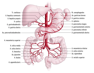
    Branches from cranial to caudal

    • Inferior phrenic arteries
    • Celiac trunk
    • Middle suprarenal artery, right and left
    • Superior mesenteric artery
    • Right or left renal artery
    • Ovarian artery - or testicular artery right and left
    • Lumbar arteries
    • Inferior mesenteric artery
    • Median sacral artery

    Supply area

    • paired branches: abdominal wall, paired retroperitoneal organs (kidneys, adrenal glands), gonads
    • unpaired branches: spleen, liver, pancreas, unpaired digestive organs
  2. Paired Branches of the Abdominal Aorta

    Main branches

    Course

    Branches and their   supply areas

    Inferior phrenic artery

    • extends cranially on the underside of the diaphragm towards the diaphragmatic domes
    • network-like division into several terminal branches

    Superior suprarenal artery: Adrenal gland

    Terminal branches: Underside of the diaphragm

    Middle suprarenal artery

    • extends laterally to the adrenal glands

    Terminal branches: Adrenal glands

    Renal artery

    • Origin approx. level L1 - 2
    • Right renal artery: runs dorsal to the inferior vena cava and pancreatic head to the right kidney
    • Left renal artery: runs dorsal to the pancreatic body to the left kidney

    Inferior suprarenal artery: Adrenal gland

    Ovarian artery, 

    Testicular artery

    • Origin below the renal arteries/L2 from the anterior side of the aorta
    • run over the psoas muscle, ureter, and external iliac arteries to the suspensory ligament of the ovary or to the deep inguinal ring of the inguinal canal and the testis

    Ovarian arteries: Ovaries, parts of the uterine tube,

    Testicular arteries: Testes, epididymides

    Lumbar arteries (bilateral 4)

    • extend laterally behind the psoas muscle over L1 – 4
    • Anastomoses with the arteries of the anterior abdominal wall (superior and inferior epigastric arteries,  iliolumbar artery, deep circumflex iliac artery)

    Spinal branches: Spinal canal

    Terminal branches: Abdominal wall, back musculature

  3. Unpaired Branches of the Abdominal Aorta

    • All unpaired branches of the abdominal aorta form anastomoses that maintain organ perfusion via collateral supply:
      • Celiac trunk → Gastroduodenal artery → Superior mesenteric artery
      • Superior mesenteric artery → Middle and left colic artery → Inferior mesenteric artery (“Riolan anastomosis”, “Drummond arcade”, rarely anastomosis according to William and Klopp)
      • Inferior mesenteric artery → Superior and middle rectal artery → Internal iliac artery
    1. Celiac trunk
    • Origin at level of T12
    • Divides into splenic artery, left gastric artery, and common hepatic artery (largest branch of the trunk)

    Main branches

    Course

    Branches and their course 

    Supply area

    Splenic artery

    • Along upper border of pancreas
    • Entry into splenorenal ligament and spleen

    Pancreatic branches:

    • Dorsal to pancreas

    Dorsal pancreatic artery:

    • Dorsal to pancreas
    • Anastomoses with great pancreatic artery to form inferior pancreatic artery

    Great pancreatic artery:

    • Dorsal to pancreatic tail caudally
    • Anastomoses with dorsal pancreatic artery to form inferior pancreatic artery

    Pancreas

    Posterior gastric artery:

    • Runs in phrenicosplenic ligament cranially to gastric fundus

    Stomach (posterior surface of fundus)

    Short gastric arteries:

    • Run in gastrosplenic ligament to gastric fundus

    Stomach (fundus and greater curvature)

    Left gastro-omental artery:

    • Runs in gastrocolic ligament to greater curvature
    • Anastomoses with right gastro-omental artery

    Stomach (fundus and greater curvature)

    Left gastric artery

    • Runs to lesser gastric curvature
    • Anastomoses with right gastric artery

    Direct branches

    Stomach (lesser curvature)

    Esophageal branches

    Esophagus

    Common hepatic artery

    • Runs to the right to hepatic portal
    • Divides into proper hepatic artery and gastroduodenal artery

    Proper hepatic artery

    • Main branch of common hepatic artery
    • Arises behind pylorus
    • Runs in hepatoduodenal ligament together with hepatic portal vein and common bile duct to hepatic portal
    • Gives off 3 branches:

    1. Right gastric artery:

    • Runs in lesser omentum to lesser gastric curvature
    • Anastomoses with left gastric artery

    Stomach (lesser curvature right)

    2. Right hepatic artery

    Liver (right part)

    Gallbladder (cystic artery)

    Partially caudate lobe

    3. Left hepatic artery

    Liver (left part)

    Partially caudate lobe

    Gastroduodenal artery

    • Runs dorsal to lesser omentum to pancreas
    • Divides into 2 branches:

    1. Right gastro-omental artery:

    • Runs to greater curvature
    • Anastomoses with left gastro-omental artery

    Pancreas, duodenum, stomach

    2. Anterior and posterior superior pancreaticoduodenal artery:

    • Form vascular plexus with branches of inferior pancreaticoduodenal artery around pancreatic head
    2. Superior mesenteric artery
    • Arises from aorta at level of L1-2
    • Runs anterior-inferior
    • Branches: Inferior pancreaticoduodenal artery, jejunal arteries and ileal arteries, ileocolic artery, right colic artery, middle colic artery

    Main branches

    Course

    Branches

    Supply area

    Inferior pancreaticoduodenal artery

    • Runs in area of horizontal part of duodenum cranially to pancreatic head

    Anterior and posterior branches:

    • Anastomose with anterior and posterior pancreaticoduodenal artery

    Pancreatic head

    Jejunal arteries and ileal arteries

    • Run in mesenteric root to jejunum and ileum
    • Form vascular arcade among themselves

    Straight arteries

    Jejunum, ileum

    Ileocolic artery

    • Runs in mesentery to ileocecal region

    Anterior and posterior cecal artery

    Cecum

    Appendicular artery (Variant: Origin from posterior cecal artery)

    Vermiform appendix

    Right colic artery

    • Runs in ascending mesocolon to ascending colon

    Divides into 2 branches, which anastomose with middle colic artery or ileocolic artery

    Ascending colon

    Middle colic artery

    • Runs in transverse mesocolon to transverse colon

    Divides into 2 branches: right branch anastomoses with right colic artery (right flexure), left branch anastomoses with left colic artery (left flexure)

    Transverse colon

    3. Inferior mesenteric artery
    • Arises at level of L3 from aorta
    • Supplies colon beyond left flexure with left colic artery, sigmoid arteries, and superior rectal artery

    Main branches

    Course

    Branches

    Supply area

    Left colic artery

    • Runs in descending mesocolon to descending colon

    Divides into 2 branches, which anastomose with middle colic artery or sigmoid arteries

    Descending colon

    Sigmoid arteries

    • 2-4 arteries that form vascular arcade in sigmoid mesocolon
    • Anastomose with left colic artery and superior rectal artery

    Several small branches to sigmoid

    Sigmoid colon

    Superior rectal artery

    • Approaches rectum from dorsal
    • Anastomoses with middle rectal artery

    Several small branches to rectum

    Upper rectum

  4. Arterial Supply of the Pelvis

    Arterial Supply of the Pelvis
    Zum Vergrößern bitte anklicken
    • Abdominal aorta divides at the aortic bifurcation (approx. level L4) into the two common iliac arteries
    • each common iliac artery divides in turn into an internal and external iliac artery
    • Internal iliac artery supplies mainly the pelvic viscera with visceral branches, with its parietal branches it is involved in the supply of the lower extremities
    • External iliac artery contributes to the supply of the pelvis and becomes the femoral artery after passing through the vascular lacuna
    1. Internal Iliac Artery

    Origin

    • arises directly caudal to the aortic bifurcation from the common iliac artery

    Course

    • passes over the upper margin of the greater sciatic foramen caudally

    Positional Relationships

    • dorsal: internal iliac vein, piriformis muscle, lumbosacral plexus
    • ventral: ureter
    • lateral: external iliac vein, psoas major muscle
    • caudal: obturator nerve

    Branches

    visceral branches:

    • Umbilical artery
    • Inferior vesical artery
    • Middle rectal artery
    • in women: uterine artery

    parietal branches:

    • Iliolumbar artery
    • Lateral sacral arteries
    • Superior gluteal artery
    • Inferior gluteal artery
    • Obturator artery
    • Internal pudendal artery

    Supply Area

    • Pelvic organs, gluteal musculature
    1.1 Visceral Branches of the Internal Iliac Artery

    Main Branches

    Course and Branches

    Supply Area

    Umbilical artery

    • postnatal obliteration to the medial umbilical ligament
    • remaining branches: superior vesical arteries, in men: artery of the ductus deferens
    • Urinary bladder body
    • in men: ductus deferens

    Inferior vesical artery

    • branching in the urinary bladder fundus into small branches
    • Urinary bladder
    • in men: prostate, seminal vesicles

    Middle rectal artery

    • branching in the middle rectal segment
    • anastomoses with superior and inferior rectal arteries
    • Rectal ampulla
    • in women: vagina
    • in men: prostate

    Uterine artery (women)

    • passes over the ureter
    • tortuous course in the broad ligament of the uterus
    • gives off numerous small branches
    • Ovary
    • Uterine tube
    • proximal vagina
    • Uterus
    1.2 Parietal Branches of the Internal Iliac Artery

    Main Branches

    Course and Branches

    Supply Area

    Iliolumbar artery

    • runs obliquely upward to the iliopsoas muscle
    • Psoas major muscle
    • Iliacus muscle

    Lateral sacral arteries

    • Superior branch: runs medially through the 1st or 2nd anterior sacral foramen and exits again through the posterior sacral foramen
    • Inferior branch: passes over the piriformis muscle and medial to the sacral foramina on the sacrum caudally
    • Sacral canal
    • Piriformis muscle
    • Muscles/skin dorsal to the sacrum

    Superior gluteal artery

    • passes with the superior gluteal nerve through the suprapiriform foramen and with branches to the gluteus maximus muscle (superficial branch) and gluteus medius muscle (deep branch)
    • Gluteus maximus, medius, and minimus muscles
    • Piriformis muscle
    • Tensor fasciae latae muscle
    • Skin over the sacrum

    Inferior gluteal artery

    • passes on the piriformis muscle caudally through the infrapiriform foramen
    • then runs together with the posterior femoral cutaneous nerve and sciatic nerve through the gluteus maximus muscle
    • Gluteus maximus, medius, and minimus muscles
    • Piriformis muscle
    • Quadriceps femoris muscle
    • Skin of the lower buttock and proximal thigh

    Obturator artery

    • passes along the lateral pelvic wall forward and downward
    • branches to the ilium and iliacus muscle
    • after passing through the obturator canal, gives off further branches, including pubic branch (anastomoses with the inferior epigastric artery to form the corona mortis) and posterior branch (branch: acetabular artery → artery of the femoral head)
    • Ilium
    • Iliacus muscle
    • Thigh adductors
    • Pubis
    • External hip muscles
    • Femoral head

    Internal pudendal artery

    • runs ventral to the sacral plexus to the infrapiriform foramen
    • re-enters the lesser pelvis via the lesser sciatic foramen, through the pudendal canal and branches in the ischiorectal fossa
    • Distal third of the rectum
    • Anal canal
    • Urethra
    • Scrotal skin/labia
    • Bulb of the penis/vestibule
    • Dorsum of the penis/clitoral prepuce
    2. External Iliac Artery

    Origin

    • arises caudal to the internal iliac artery from the common iliac artery

    Course

    • runs along the psoas major muscle to the inguinal ligament, through the vascular lacuna and becomes the femoral artery here

    Positional Relationships

    • Right external iliac artery runs under the ureter

    Branches

    • Deep circumflex iliac artery runs to the anterior superior iliac spine and anastomoses with the iliolumbar artery and superior gluteal artery
    • Inferior epigastric artery runs medial to the deep inguinal ring cranially and between the transversus abdominis muscle and rectus abdominis muscle, further branches including cremasteric artery or round ligament artery 

    Supply Area

    • Ilium, lower portion of the abdominal wall
  5. Femoral artery

    Femoral artery
    Zum Vergrößern bitte anklicken
     1. Overview

    Origin

    • External iliac artery, which becomes the femoral artery after passing under the inguinal ligament

    Course

    • runs between M. iliopsoas and M. pectineus (= iliopectineal fossa) covered by fascia lata
    • is accompanied by M. sartorius up to the middle of the thigh
    • passes together with V. femoralis and N. saphenus through the adductor canal
    • transitions into the popliteal artery after passing through the adductor hiatus

    Branches

    • A. epigastrica superficialis
    • A. circumflexa ilium superficialis
    • A. profunda femoris (main artery for supplying the thigh!)
    • Aa. pudendae externae
    • A. descendens genus

    Supply area

    • Thigh 
    • Skin of the abdominal wall
    • External genitalia
    • Knee joint and proximal/medial lower leg
    2. Important branches of the femoral artery

    Branches

    Supply area

    A. epigastrica superficialis

    • Ø
    • Skin of the abdominal wall up to the umbilical region

    A. circumflexa ilium superficialis

    • Ø
    • Anterior superior iliac spine

    A. profunda femoris

    • A. circumflexa femoris medialis runs between M. iliopsoas and M. pectineus dorsally → trochanteric fossa; anastomosis with A. circumflexa femoris lateralis
    • A. circumflexa femoris lateralis runs between M. rectus femoris and Mm. vasti laterally; anastomosis with A. circumflexa femoris medialis
    • Aa. perforantes pass through the adductors to the back of the thigh
    • Thigh

    Aa. pudendae externae

    • Ø
    • External genitalia
    • Scrotum or labia majora

    A. descendens genus

    • Rr. articulares → Rete articulare genus
    • R. saphenus → runs medially together with V. saphena magna and N. saphenus to the calf
    • Knee
    • Proximal medial calf