Start your free 3-day trial — no credit card required, full access included

Perioperative management - Endovascular bifurcation prosthesis for infrarenal AAA with simultaneous aneurysm of the right common iliac artery (EVAR with iliac sidebranch)

  1. Indications

    • Abdominal aortic aneurysm (AAA) > 5 cm
    • smaller aneurysms in eccentric AAA or ulcerations with contained perforation (PAU, penetrating aortic ulcer)
    • symptomatic aneurysms of any size

    Special indications and therapy options for Behçet's disease, Marfan syndrome, mycotic aneurysms such as e.g. Salmonella infections and AAA with peripheral embolization.

    The indication for invasive therapy of an abdominal aortic aneurysm (AAA) basically results from a comparison of the individual rupture risk in the natural course against the surgical risk. If the rupture risk in the natural course exceeds the individual surgical risk, the indication for invasive therapy is generally given.

    Classification of Rupture Risk

    Factors

    Low Risk

    Medium Risk

    High Risk

    Aneurysm diameter

    < 5 cm

    5-6 cm

    > 6 cm

    Growth rate per year

    < 0,3 cm

    0,3-0,5 cm

    > 0,5 cm

    Smoking/COPD

    low

    moderate

    high

    Family history

    none

    isolated

    frequent

    Arterial hypertension

    none

    well controlled

    unstable despite therapy

    Morphology

    fusiform

    saccular

    bulges

    Gender

    male

    female

    Surgical Indication

    Classification

    Size

    Wall

    Clinical presentation

    Surgical Indication

    asymptomatic infrarenal

    > 5 cm  ♂

    > 4,5 cm  ♀

    intact

    none

    elective

    asymptomatic suprarenal

     > 6 cm

    intact

    none

    elective

    symptomatic

    independent

    intact

    Spontaneous pain, tender abdomen, back or flank

    urgent, within 24 hrs.

    ruptured

    independent

    contained or free ruptured

    diffuse severe spontaneous/touch pain of the tense abdomen, with/without hemorrhagic shock

    Emergency

    aortoduodenal fistula

    intermittent vomiting, melena

    Emergency

    aortocaval fistula

    Right heart failure, fistula sounds, truncal cyanosis, simultaneous contrast enhancement aorta & inferior vena cava

    Emergency

    For the surgical treatment of the AAA, two procedures are available:

    • the open replacement of the abdominal aorta with a tube or bifurcation prosthesis (OAR, open aortic repair)
    • the endovascular implantation of a stent prosthesis (EVAR, endovascular aortic repair)

    Laparoscopic aneurysm surgery, usually in combination with a mini-laparotomy, is of rather minor importance.

    For the selection of the procedure - OAR or EVAR - the following recommendations exist:

    OAR (trans-, retroperitoneal)

    • normal life expectancy
    • low surgical risk ( “fitness”)
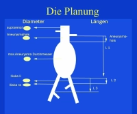
    • unsuitable anatomy for EVAR: landing zone, aneurysm neck (angle, length), iliac vessels (stenoses, elongation, kinking), thrombi, calcification
    • Marfan and other connective tissue diseases

    EVAR (standard prosthesis, custom-made)

    • previous abdominal operations
    • limited life expectancy
    • high surgical risk
    • anatomy suitable for EVAR (see above)

    EVAR requires adequate iliac vessels for access, as the stent graft systems are often large-caliber. Problematic are atherosclerotically narrowed, tortuous and kinked, but also aneurysmatically dilated iliac vessels.

    In the long-term course, the endovascular aortic prosthesis is associated with a higher complication rate than open aortic surgery.

    The mortality risk for EVAR or OAR of an individual patient can be quickly calculated using the so-called BAR Score Calculator → www.britishaneurysmrepairscore.com, which can be usefully used for patient counseling about the risk of an elective procedure with EVAR or OAR.

    The general surgical indication is given for (isolated) iliac aneurysm from an aneurysm diameter of 3 cm.

    Video example:

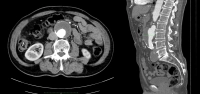
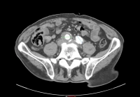
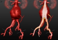
    • infrarenal AAA, diameter 54.2 mm
    • aneurysm of the right common iliac artery, diameter 41.1 mm
    PM 327-1
    Abb. 1 BAA transversal/sagital
    PM 327-2
    Abb. 2 Iliakalaneurysma transversal
    PM 327-3
    Abb. 3 Aortoiliakales Aneurysma rechts 3D, re. Bild mit Lumen; Duplikatur re. Nierenarterie
  2. Contraindications

    Because in interventional procedures, due to unexpected technical problems, a change in procedure to an open operation may become necessary, the preoperative risk assessment and the definition of contraindications are important:

    • Heart failure (ejection fraction < 25-30 %)
    • COPD (FEV1 < 0,8)
    • non-correctable severe CAD
    • incurable cancer

    Relative contraindication:

    • compensated renal insufficiency (due to intraoperative contrast medium load)

    If in borderline cases the operation is to be performed despite contraindications, this requires special agreement with the patient or with his relatives.    

    Technical contraindications for endovascular treatment: see Preoperative Diagnostics/Planning

  3. Preoperative Diagnostics

    Thorough vascular surgical examination:

    • Pulse status
    • Doppler legs
    • if necessary, walking distance test in case of concurrent PAD and poorly palpable foot pulses
    • Abdominal ultrasound
    • Color duplex of carotids
    • Echocardiography
    • Stress ECG
    • Laboratory tests (electrolytes, coagulation, renal values, blood count, blood lipids)
    • Chest X-ray
    • Spiral CT
    • if necessary, in case of concurrent PAD or critical visceral artery morphology i.a. DSA of the abdominal aorta and pelvic and leg vessels

    Important is especially the preoperative angiographic clarification of the access pathology:

    • Stenoses of the femoral and pelvic arteries
    • Aneurysms of the access arteries
    • local infections such as e.g. erythrasma
    • Previous operations in the surgical area

    A special feature is the presence of a horseshoe kidney in AAA, where both in planned open procedure and in endovascular procedure a special planning is necessary, which must take into account the preservation of the multiple renal arteries in the area of the abdominal aortic aneurysm.

    In summary, the preoperative diagnostics must therefore consider the following parameters:

    1. Aortic pathology

    2. Risk factors (multimorbidity)

    3. Access pathology

    Planning

    In this context, the measurement of the aneurysm and the consideration of calcifications and stenoses are of decisive importance to check the local feasibility.

    PM 327-4
    Abb. 4 Präoperative Vermessung des BAA

    One of the possible contraindications to endovascular treatment is severe kinking of the aneurysm neck. Furthermore, coiling, compression and elongation must be considered, because the prosthesis - depending on the centerline - may deviate from the aneurysm configuration and it can subsequently lead to misplacements.

    When measuring, the exact guidelines of the prosthesis manufacturer must be observed. These differ depending on the prosthesis type. For example, an oversizing of the iliac prosthesis between 7 and 35 % should be performed. Each manufacturer has its own case planning forms and its particular guidelines regarding minimum lengths, -diameters, oversizing, minimum overlaps etc. Also, the recommended accessories can vary depending on the prosthesis manufacturer and must be strictly observed.

  4. Special Preparation

    • Determine blood type, if necessary provide blood products
    • Depilate the surgical area
    • if necessary, insert urinary catheter
    • Prophylactic antibiosis is usually performed and is recommended by the German Society for Vascular Surgery (single-shot Cefuroxime 30 minutes before skin incision), however, the benefit is currently being discussed due to the resistance issues (RKI).  
  5. Informed Consent

    General Surgical Risks

    • Severe bleeding, blood transfusions, transmission of Hepatitis/HIV through donor blood products
    • Allergy/Intolerance
    • Wound infection
    • Thrombosis/Embolism
    • Skin, vascular, nerve damage e.g. due to positioning
    • Keloids (if open access)

    Specific Procedure Risks

    • Vascular injuries e.g. during prosthesis placement: surgical hemostasis, blood transfusions, in case of severe bleeding or aortic tear immediate conversion to open surgery
    • Pseudoaneurysm of the punctured vessels, AV fistula, seroma
    • Primary aortoenteric fistula: Surgical intervention, possibly with removal of the vascular prosthesis
    • Inadequate anchoring or leakage of the prosthesis: Corrective procedures, possibly open
    • Infection of the vascular prosthesis: possible after days, months or years; Endocarditis, sepsis, leg ischemia, amputation; surgical removal of the vascular prosthesis
    • Damage to adjacent organs; Intestinal ischemia → Resection, stoma placement
    • Nerve lesions → Sensory disturbances, pain, paralysis of the abdominal wall, thigh muscles
    • Lymph fistula
    • Lymphedema of the legs; temporary, permanent; compression stockings, lymphatic drainage
    • Postoperative bleeding
    • Impotence    
    • Compromised kidney function due to intraoperative angiography, permanent dialysis

    Risks from Perfusion Disorders

    • Thrombosis/Embolism: Pulmonary embolism, stroke, myocardial infarction; Prophylaxis with heparin → HIT II risk
    • Legs: Thrombosis of the vascular prosthesis and possibly adjacent vascular segments, possibly leg ischemia, amputation (e.g. toes)
    • Kidneys: due to thrombosis or placement of the vascular prosthesis; Despite immediate surgical correction, possibly permanent kidney damage → dialysis
    • Intestine: Perfusion disorders due to thrombosis/embolism and possibly placement of the vascular prosthesis; vascular surgical intervention required, possibly intestinal resection, permanent stoma
    • Spinal cord: depending on the size of the aneurysm, perfusion disorders due to placement of the vascular prosthesis; temporary sensory disturbances/paresis, possibly also permanent paraplegia in case of low-originating A. radicularis magna
    • Gluteal muscles: due to overstenting of the pelvic arteries, especially bilaterally; Claudication symptoms when walking, possibly gluteal necrosis formation
    • Liver: Functional impairment due to hypoperfusion, especially in pre-existing liver conditions
  6. Anesthesia

    • ITN
    • in suitable patients (compliance) also local anesthesia
  7. Positioning

    Positioning

    Supine position, both arms abducted. The upper body should be slightly reclined at the level of the thoracoabdominal junction between the pubic symphysis and xiphoid process. Pack both feet in cotton boots (Caution pressure injury).

  8. OR Setup

    OR Setup

    In the video example, the surgeon stands on the right; the side is only briefly switched for the exposure of the left femoral artery. The surgical assistant stands on the left side at the foot end of the patient. The DSA unit incl. monitor is also located on the left.

  9. Special Instrumentation and Holding Systems

    Special Instrumentation and Holding Systems
    Prothesen-Module
    • small vessel sieve
    • sufficient guidewires (0.035´´ soft guidewire e.g. Terumo®, 0.035´´ superstiff guidewires e.g. Lunderquist®, Amplatz®, Backup Meyer®)
    • guide catheters and selective angiography catheters with different configurations (e.g. Hockey Stick®, Renal Double Curve®, Sidewinder® etc.)
    • dilatation balloons in various sizes (6-9 mm)
    • stents for renal arteries and for additional fixation of the prosthesis if necessary
    • long and short pigtail catheters, pigtail with graduation
    • 16 & 18 Fr. introducer sheath, 12 Fr. flexible sheath 45 cm (all sheaths with hydrophilic coating)
    • aortic molding balloon 16 – 34 mm
    • PTA balloon 10 – 14 mm (compatible for guidewire 0.035´´, balloon length 4 cm, usable length 75 cm)
    • 0.035´´ superstiff pull-through wire
    • snare catheter sets
    • balloon- and self-expandable stents fitting all diameters of the endoluminal prosthesis
    • if necessary, embolization coils, percutaneous vascular closure systems
    • DSA system, contrast medium high-pressure pump
    • suitable endoluminal vascular prostheses according to preoperative measurement
    • reserve materials for central and peripheral prosthesis extension
    • keep instrumentation for open conversion ready in case of emergency
  10. Postoperative Treatment

    Postoperative Analgesia

    Follow the link here to PROSPECT (Procedure Specific Postoperative Pain Management) and to the current guideline Treatment of acute perioperative and posttraumatic pain.

    Medical Follow-up Care

    • 24-hour follow-up care in intensive care unit, possibly intermediate care
    • Normal ward possible from the 1st postoperative day
    • Close monitoring of heart-circulation-lungs
    • Coagulation monitoring (e.g., consumption coagulopathy)
    • Check pulses in the legs, capillary perfusion of the feet

    Thrombosis Prophylaxis

    • Weight-adapted low-molecular-weight heparin; in multifocal atherosclerosis ASA 100 mg once daily starting with food intake
    • For Marcumar® patients, switch from heparin to Marcumar® on the 5th postoperative day

     Mobilization

    • From 1st/2nd postoperative day to bed edge

    Physiotherapy

    • Isometric muscle training, breathing exercises    

    Diet Build-up

    • 4 – 6 hours postoperatively

    Bowel Regulation

    • On the 3rd day enema if no spontaneous bowel movement has occurred

    Incapacity for Work

    • approx. 3 months