Start your free 3-day trial — no credit card required, full access included

Anatomy - Femoropopliteal bypass on the right with PTFE prosthesis (PIII)

  1. Arterial blood supply to the lower extremity

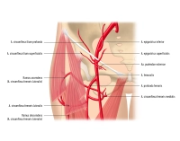
    1. A. femoralis

    A 311-1
    Zum Vergrößern bitte anklicken
    1.1. Overview

    Origin

    • A. iliaca externa, which becomes the A. femoralis after passing beneath the inguinal ligament

    Course

    • runs between the M. iliopsoas and M. pectineus (= fossa iliopectinea), covered by the fascia lata
    • is accompanied by the M. sartorius as far as the mid-thigh
    • passes together with the V. femoralis and the N. saphenus through the adductor canal
    • continues as the A. poplitea after passing through the hiatus adductorius

    Branches

    • A. epigastrica superficialis
    • A. circumflexa ilium superficialis
    • A. profunda femoris (main artery supplying the thigh!)
    • Aa. pudendae externae
    • A. descendens genus

    Area of supply

    • Thigh
    • Skin of the abdominal wall
    • External genitalia
    • Knee joint and proximal/medial lower leg
    1.2. Important branches of the A. femoralis

    Branches

    Area of supply

    A. epigastrica superficialis

    • Ø
    • Skin of the abdominal wall up to the umbilical region

    A. circumflexa ilium superficialis

    • Ø
    • Spina iliaca anterior superior

    A. profunda femoris

    • A. circumflexa femoris medialis passes dorsally between the M. iliopsoas and M. pectineus → fossa trochanterica; anastomoses with the A. circumflexa femoris lateralis
    • A. circumflexa femoris lateralis passes laterally between the M. rectus femoris and the Mm. vasti; anastomoses with the A. circumflexa femoris medialis
    • Aa. perforantes pass through the adductors to the posterior aspect of the thigh
    • Thigh

    Aa. pudendae externae

    • Ø
    • External genitalia
    • Scrotum or labia majora pudendi

    A. descendens genus

    • Rr. articulares → rete articulare genus
    • R. saphenus → passes medially together with the V. saphena magna and the N. saphenus to the calf
    • Knee
    • Proximal medial calf

    2. A. poplitea

    A 311-2
    Zum Vergrößern bitte anklicken
    2.1. Overview

    Origin

    • Continuation of the A. femoralis after its passage through the hiatus adductorius

    Course

    • passes through the popliteal fossa
    • runs between the heads of the M. gastrocnemius
    • continues as the truncus tibiofibularis after giving off the A. tibialis anterior

    Branches

    • A. tibialis anterior

    Area of supply

    • Popliteal fossa
    • Knee joint
    2.2. Important branches of the A. poplitea

    Course

    Branches

    Area of supply

    A. tibialis anterior

    • Branch of the A. poplitea
    • is accompanied by 2 veins and, in its lower part, by the N. peronaeus profundus
    • passes through the membrana interossea cruris and runs on its anterior aspect
    • runs behind the tendon of the M. extensor hallucis longus
    • continues as the A. dorsalis pedis
    • A. recurrens tibialis posterior + anterior
    • A. malleolaris anterior lateralis + medialis
    • A. dorsalis pedis (terminal branch of the A. tibialis anterior)
    • Anterior lower leg
    • Dorsum of the foot

    From a surgical-technical perspective, it is customary to divide the A. poplitea into three segments (PI – PIII), which guide the operative approaches:

    Segment

    Feature

    Approach

    PI

    From the exit of the adductor canal to the gastrocnemius tunnel proximal to the knee joint

    Distal medial thigh

    PII

    Knee joint

    Posterior (popliteal fossa)

    PIII

    From the exit of the gastrocnemius tunnel below the knee joint to the origin of the A. tibialis anterior/soleus arch

    Proximal medial lower leg

    The A. poplitea runs within a neurovascular bundle surrounded by fat-rich connective tissue (corpus adiposum popliteum). In the PI and PII segments the artery lies ventromedial to the V. poplitea, while in the PIII segment it is surrounded by the Vv. comitantes. The N. tibialis and the N. peroneus communis lie dorsolateral to the A. and V. poplitea. In the popliteal fossa the nerves have a relatively superficial position, which must be taken into account when dissecting the A. poplitea from the dorsal aspect.

    The superficial and deep lymphatic drainage in the fossa poplitea is bundled together and is drained via three to five perivenously located lymph nodes.

    Owing to its location, the A. poplitea is subjected to considerable mechanical stress, which is why, as an artery of the muscular type, it possesses strong muscular and fibrous layers (tunica media, tunica elastica interna). Its wall properties are therefore similar to those of the central elastic arteries.

    3. A. tibialis posterior

    3.1. Overview

    Origin

    • Continuation of the A. poplitea after the origin of the A. tibialis anterior

    Course

    • passes together with the N. tibialis beneath the tendinous arch of the M. soleus → sulcus malleolaris medialis → malleolus medialis
    • Division into the A. plantaris medialis + lateralis

    Branches

    • A. fibularis
    • A. plantaris medialis + lateralis

    Area of supply

    • Posterior aspect of the lower leg
    • Sole of the foot
    3.2. Important branches of the A. tibialis posterior

    Course

    Branches

    Area of supply

    A. fibularis

    • runs along the posterior aspect of the fibula
    • runs between the M. tibialis posterior and the M. flexor hallucis longus
    • R. communicans: transverse connection to the A. tibialis posterior
    • Rr. malleolares laterales
    • Lateral-dorsal lower leg

    A. plantaris medialis

    • runs in the medial neurovascular strand of the sole (A., V. and N. plantaris medialis) between the M. abductor hallucis and the M. flexor digitorum brevis
    • Ø
    • Medial sole of the foot

    A. plantaris lateralis

    • runs in the lateral neurovascular strand of the sole (A., V. and N. plantaris lateralis) between the M. flexor digitorum brevis and the M. quadratus plantae
    • Arcus plantaris profundus: anastomosis between the A. plantaris lateralis and the R. plantaris profundus of the A. dorsalis pedis in the region of the metatarsal bones
    • Lateral sole of the foot
    • Toes
to top