Start your free 3-day trial β€” no credit card required, full access included

Perioperative management - Femoropopliteal bypass on the right with PTFE prosthesis (PIII)

  1. Indications

    The TASC II (Transatlantic Inter-Society Consensus for the Management of Peripheral Arterial Disease) consensus document addresses aspects of revascularisation in PAOD.

    According to the TASC criteria, therapeutic treatment options can be derived depending on the length of the local occlusion/stenosis. The length of the stenosis and its regional location determine the treatment: endovascular or open vascular surgery.

    TASC criteria for femoropopliteal occlusions

    TypeMorphologyProcedure
    ASingle stenosis <5 cm in length, not at the origin of the SFA or in the distal popliteal artery; single occlusion <3 cm in length (not at the origin of the SFA or popliteal artery)Endovascular
    BSingle stenosis 5–10 cm in length, not in the distal popliteal artery; single occlusion 3–10 cm in length, not in the distal popliteal artery; calcified stenosis <5 cm in length; multiple lesions <3 cm in lengthEndovascular
    CSingle occlusion 3–10 cm in length extending to the distal popliteal artery; multiple focal lesions 3–5 cm in length without/with calcification; single stenosis/occlusion >10 cm in lengthOpen revascularisation
    DComplete occlusion of the CFA and/or SFA; complete occlusion of the popliteal artery and trifurcation; severe diffuse diseaseOpen revascularisation

    Video example: TASC D → open revascularisation

    Right leg PAOD Fontaine grade IV, left leg grade III with:

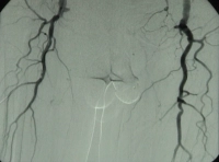
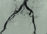
    • High-grade arteriosclerosis of the terminal aorta and both iliac arteries
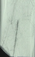
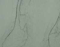
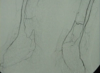
    • Extended occlusion of both superficial femoral arteries
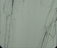
    • Partial occlusion of the infrapopliteal arteries in both legs

    Preoperative DSA of the patient:
    PM 311-1
    Figure 1: High-grade arteriosclerosis of the terminal aorta and both iliac arteries


    PM 311-2
    Figure 2: Extended occlusion of both superficial femoral arteries


    PM 311-3
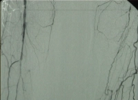
    Figure 3: Filling of popliteal segment P1 via collaterals


    PM 311-4
    Figure 4: Partial occlusion of the infrapopliteal arteries in both legs, occlusion of popliteal segment P3 on the left


    PM 311-5
    Figure 5: Partial occlusion of the infrapopliteal arteries in both legs


    PM 311-6
    Figure 6: Occlusion of the right posterior tibial artery


    PM 311-7
    Figure 7: Both plantar arches without visualization
  2. Contraindications

    • Infection in the region of the repair
    • ASA IV
    • Previous inguinal radiotherapy (which would then mandate extra-anatomical bypass routing)
  3. Preoperative diagnostic work-up

    Medical history

    • Claudication
    • Walking distance
    • Risk factors -> nicotine abuse; arterial hypertension; coronary heart disease; cardiac failure; diabetes mellitus; manifest renal failure with/without dialysis dependence; coagulopathies

    Inspection

    • Skin changes
    • Muscular abnormalities
    • Orthopaedic malalignment
    • Skin colour
    • Body hair
    • Trophic changes
    • Swelling; oedema; mycosis; phlegmon; ulcera cruris, etc.

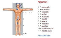
    Palpatory bilateral comparison

    • Pulse status
    • Skin temperature

    Auscultatory bilateral comparison of the limb arteries

    Palpation-Auskultation
    Palpation–auscultation

    Ankle-Brachial Index (ABI)

    • ABI = systolic BP of posterior tibial artery / systolic BP of brachial artery
    ABI valuePAOD severity
    > 1.3Falsely high values (suspected Mönckeberg medial sclerosis, e.g. in diabetes mellitus)
    > 0.9Normal finding
    0.75 - 0.9Mild PAOD
    0.5 - 0.75Moderate PAOD
    < 0.5Severe PAOD
    • An ABI value of < 0.9 is considered evidence of significant PAOD.
    • Determining the ankle-brachial index (ABI) through non-invasive Doppler occlusion pressure measurement is a suitable test for confirming PAOD.
    • PAD diagnosis is determined by the ABI value with the lowest ankle artery pressure.
    • A pathological ankle-brachial index is an independent risk indicator for increased cardiovascular morbidity and mortality.

    Colour-coded duplex ultrasonography

    • Carotid arteries, abdominal aorta, limb arteries
    • Localisation of stenoses and occlusions in almost all vascular regions apart from the thoracic region
    • Allows quantification of the degree of stenosis and assessment of plaque morphology
    • Sensitivity and specificity around 90%
    • Well suited as a screening modality

    CT angiography

    • Multislice computed tomography (MS-CT) with nonionic contrast agent
    • Broad range of indications: traumatic vascular lesion (esp. trunk); vascular dissection/rupture; aneurysm; arterial thrombosis/embolism; portal vein/mesenteric vein thrombosis; pulmonary artery embolism; PAD; vascular tumours
    • Benefits: rapid; detects relevant comorbidities; visualises peripheral arteries; sensitivity and specificity each about 90%
    • Drawbacks: radiation and contrast agent exposure, allergies (about 3%), no functional assessment

    Cardiac check

    • Resting ECG
    • Exercise ECG
    • Echocardiography

    Chest X-ray

    Possibly spirometry

    Laboratory panels

    • Blood count
    • Electrolytes
    • Coagulation
    • Kidney function parameters
    • Liver function parameters
    • Blood lipids
    • Blood group
    • In PAOD Fontaine grade IV → wound swab/antimicrobial susceptibility testing
  4. Special preparation

    • Trimming the hair in the surgical field
    • Order packed RBCs
    • Protect the foot of the side to be operated on with a cotton-wool shoe (to avoid intraoperative pressure injury when clamping the vessels)
    • Antibiotic therapy according to antibiogram (see diagnostic work-up) for 5 days postoperatively, depending on local findings
  5. Informed consent

    General surgical risks

    • Major bleeding, blood transfusions, transmission of hepatitis/HIV through allogeneic blood transfusions
    • Allergy/intolerance
    • Wound infection
    • Thrombosis/embolism
    • Skin, vascular and nerve damage, e.g., due to patient positioning
    • Keloids

    Specific procedural risks

    • Thrombotic occlusion of the bypass and possibly the adjacent vessel segment; operative revision, (partial) amputation
    • Massive infection with severe bleeding from the bypass suture lines; bypass explantation, (partial) amputation, sepsis
    • Injury to sensory and motor nerves; dysaesthesia, pain, temporary or even persistent (partial) palsy of the thigh muscles
    • Injury to lymph vessels; temporary or persistent lymphoedema, lymphatic fistula
    • Compartment syndrome; possibly requiring surgical pressure relief/fasciotomy, persistent palsy, limb loss
    • Renal dysfunction with temporary or permanent dialysis due to tourniquet syndrome and intraoperative angiography (contrast medium)
    • Suture line aneurysm; surgical intervention depending on size/presentation
Anaesthesia

General anaesthesiaSpinal anaesthesia (Caution: the patient may not notice new-onset ischaemia in e

Activate now and continue learning straight away.

Single Access

Activation of this course for 3 days.

US$9.30  inclusive VAT

Most popular offer

webop - Savings Flex

Combine our learning modules flexibly and save up to 50%.

from US$4.30 / module

US$51.61/ yearly payment

price overview

vascular surgery

Unlock all courses in this module.

US$8.60 / month

US$103.20 / yearly payment

to top