Start your free 3-day trial — no credit card required, full access included

Perioperative management - Alloplastic carotid-subclavian bypass for left subclavian artery stenosis

  1. Indications

    Indications

    Stenosis or occlusion of the subclavian artery with clearly attributable symptoms:

    • clinically relevant vertebrobasilar insufficiency that is reproducible by provocation tests
    • hypoperfusion of the arm requiring treatment (brachial claudication)
  2. Contraindications

    • general inoperability (severe CHD, COPD, inoperable tumor disease)
    • inoperable peripheral vasculature
    • Status post radiation in the vascular region, e.g., after neck dissection
    • asymptomatic stenoses!
  3. Preoperative Diagnostics

    History

    • vascular risk factors: smoking, arterial hypertension, lipid metabolism disorders, diabetes mellitus

    Clinical-Neurological Examination

    • > 90 % of stenoses and occlusions of supra-aortic vessels (ICA, vertebral artery, etc.) remain clinically asymptomatic and are discovered incidentally during screening examinations or preoperative imaging
    • the symptomatology derives from the location of the lesions at the distribution point of cerebral perfusion and the supply of the upper extremity

    1. Cerebral Symptoms

    • occlusive processes of the subclavian artery near the aortic arch before the origin of the vertebral artery → vertebrobasilar insufficiency: intermittent vertigo ("drop-attacks"), inner ear hearing loss, gait instability
    • occlusive processes in the vertebral artery territory -> Wallenberg syndrome: Horner syndrome, ataxia, velar paresis (each ipsilateral), dissociated sensory disturbances contralateral
    • Steal phenomenon: provocation of symptoms (vertigo, drop-attacks) by working with the arm on the affected side (due to withdrawal of blood from the basilar territory via retrograde flow of the vertebral artery)

    2. Peripheral Symptoms

    • blood pressure reduction in the affected arm
    • weakened or absent pulse in the upper extremity
    • brachial claudication

    Doppler and Duplex Sonography

    In the ultrasound examination of the extracranial brain-supplying vessels, all vessels should always be assessed in transverse and longitudinal sections:

    • common carotid artery from proximal to the carotid bifurcation
    • carotid bifurcation with dorsolateral originating internal carotid artery
    • external carotid artery
    • vertebral artery in segments V1 to V3
    • subclavian artery and axillary artery

    Both the brachiocephalic trunk and the origin area of the left subclavian artery are usually not directly visible for anatomical reasons. However, from the flow spectrum of the more cranial arterial sections, stenoses in the inflow can be indirectly inferred (fist closure test, application of a blood pressure cuff → flow reversal).

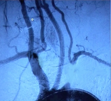
    Digital Subtraction Angiography (DSA), MR Angiography

    • depiction of the supra-aortic circulation including arm and the steal circuit

    CT or MRI Brain

    • exclusion of cerebral ischemic events

    Cardiological Examination

    • resting, stress ECG
    • if necessary, cardiac echo

    Chest X-ray Examination

    Laboratory

    • CBC
    • electrolytes
    • coagulation
    • retention values
    • liver enzymes
    • blood lipids
    • blood group
  4. Special Preparation

    • Mark the side, shave
    • Fasting 2 – 6 hours depending on clinic standard
    • Leave platelet aggregation inhibitors; in dual therapy, individual decision depending on cardiac risk profile
  5. Informed Consent

    General Surgical Risks

    • Severe bleeding, blood transfusions, transmission of Hepatitis/HIV through blood products
    • Allergy/Intolerance
    • Wound infection
    • Thrombosis/Embolism
    • Skin, vascular, nerve damage e.g. due to positioning
    • Keloids

    Specific Surgical Risks

    • Cerebral perfusion disorders/Stroke
    • Injury to neck nerves: Hoarseness, voice loss, swallowing and breathing disorders, mouth corner paresis, uncontrolled tongue movements, shoulder elevation weakness, Horner's syndrome (Miosis, Ptosis, Enophthalmos)
    • Postoperative bleeding, possibly emergency surgical relief
    • Increased infection risk with vascular prostheses made of plastic; Sepsis, possibly surgical revision with removal of the prosthesis
    • Cardiopulmonary complications: Myocardial infarction, Pneumonia, Pulmonary embolism
    • Suture aneurysm -> Re-OP
    • Intraoperative angiography: contrast medium-induced renal insufficiency
    • Lymph fistula, Lymph cyst
  6. Anesthesia

    ITN

  7. Positioning

    Positioning
    • Supine position with slightly elevated upper body or Beach-Chair position
    • the head rests in a head cup or on a silicone ring, is turned toward the contralateral side and slightly hyperextended
    • the arm on the operative side is adducted, on the contralateral side possibly abducted
    • Underpadding of the ipsilateral shoulder
  8. OR Setup

    OR Setup
    • Surgeon stands on the side to be operated on at shoulder height
    • 1st Assistant opposite Surgeon
    • Instrumenting OR staff towards the feet next to 1st Assistant
    • If necessary, 2nd Assistant on the side to be operated on at the patient's head height
  9. Special Instrument Sets and Holding Systems

    • small vascular instrument set
    • blunt wound retractors
    • suction device
    • mobile DSA unit
    • monofilament suture material 5-0 to 7-0
    • Vessel loops
    • vascular prosthesis 
  10. Postoperative Treatment

    Postoperative Analgesia

    Follow the link here to PROSPECT (Procedure Specific Postoperative Pain Management) and to the current Guideline for the Treatment of Acute Perioperative and Posttraumatic Pain.

    Medical Follow-up Care

    • Monitoring in Intermediate Care for 24 hours
    • Inspection of the surgical site: Wound area and drainage
    • Circulatory monitoring
    • Laboratory checks
    • Weight-adapted heparin overlapping with platelet aggregation inhibitors
    • For arrhythmias/valvular defects, coumarin

    Mobilization

    • 1st postoperative day

    Physiotherapy

    • generally dispensable

    Diet Build-up

    • 6 hours postoperatively

    Bowel Regulation

    • generally dispensable

    Inability to Work

    • 3-4 weeks