Thoracic outlet syndrome (TOS) - Left transaxillary first rib resection (TFRR) - Vascular surgery

Reading time readingtime 18:12 min.
Activate now
  1. Axillary approach to the left first rib

    Video
    Axillary approach to the left first rib

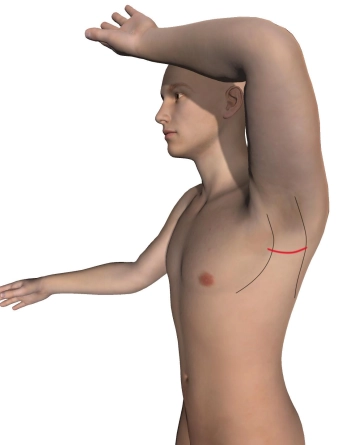
    Make a horizontal, curved skin incision in the left axilla over the palpated third rib (usually at the inferior border of the hairline) from the lateral aspect of the pectoralis major muscle to the anterior aspect of the latissimus dorsi muscle. Carry the partly sharp, partly blunt dissection down to the chest wall. Transect the longitudinal lymphatics between ligatures and bluntly sweep away the axillary lymphatic and adipose tissue until the thoracodorsal nerve and long thoracic nerve become visible, as well as the intercostobrachial nerve, which travels from the second intercostal space into the axillary adipose tissue.

    Divide between ligatures the lateral thoracic artery and the thoracoepigastric vein, the accompanying vessels of the latter nerve. Insert bladder retractors anteriad at the pectoralis major muscle and posteriad at the latissimus dorsi muscle.

    Tips:

    1. The transaxillary approach is well suited to clear the first rib with rather good visibility and allows for any needed additional intraoperative procedures (arterial repair, venous thrombectomy and thoracic sympathectomy). Wound healing and the cosmetic outcome of the scar located in the hairy axilla, are generally considered to be very good.

    2. If the skin incision is too far superior, access through the adipose and lymphatic tissue of the axilla becomes more difficult.

    3. All nerves must be spared and left in place on the chest wall. In other words, do not elevate them with the muscles!


  2. Exposing the anatomic structures superior to the first rib

    Video
    Exposing the anatomic structures superior to the first rib

    After ligating and dividing the intercostal vessels, the anterior aspect of the first rib is exposed. Raising the arm will dimly reveal the anatomic structures of the scalene gaps:

    • The tendon of the subclavian muscle (= pectoralis minor muscle) is attached to the first rib anterior to the subclavian vein
    • The anterior scalene muscle lies between the subclavian vein and artery
    • Posterior to the subclavian artery runs the brachial plexus, which is bordered posteriad by the middle scalene muscle

     Tips:

    1. The posterior scalene muscle is located more posteriad and does not need to be exposed or divided in most cases.

    2. Sometimes a scalenus minimus muscle traveling to the first rib is found between the subclavian artery and the brachial plexus, which must then be carefully divided without injuring the subclavian artery.

    3. After each surgical step, relieve the traction on the arm to avoid brachial plexus injury.


  3. Exposing the tendon of the subclavian muscle and the anterior scalene muscle

    Video
    Exposing the tendon of the subclavian muscle and the anterior scalene muscle

    The video clip once again shows the anatomic structures and their spatial relationship: Subclavian artery and vein, brachial plexus, subclavian muscle, and anterior scalene ("anticus") muscle.

    Tips:

    1. Careful dissection is needed here to avoid injury to the subclavian vein.  It is difficult to control major bleeding here at this stage of the dissection.

    2. The costoclavicular ligament is exposed beneath the transected tendon. Leave this ligament untouched!

  4. Transecting the anterior scalene muscle

    Video
    Transecting the anterior scalene muscle

    Bluntly expose the scalenus anterior muscle between the subclavian artery and vein, gradually free it with Overholt forceps at the anterior and posterior aspect, and finally tunnel the muscle with the forceps. Have the assistant gently raise the arm and move it anteriad or posteriad, depending on the view of the scalene gap. Completely transect the muscle around 1 cm superior to the first rib, step by step, either with a long scalpel or with scissors, as demonstrated in the video clip.

    Tips:

    1. Careful dissection is needed here to avoid injury to the subclavian vein. It is difficult to control major bleeding here at this stage of the dissection.

    2. The pleura may be injured during dissection and tunneling of the scalene anterior muscle with the Overholt forceps. Small pleural defects are not a problem and can be controlled at the end of the operation with a chest tube. But with larger defects the collapsed lung will impede dissection.

    3. In case of pleural defects, inform the anesthetist -> possibly double lumen tube, change in ventilation mode, etc.


Transecting the middle scalene muscle

Tunnel the middle scalene muscle with the Overholt forceps and divide the muscle at the upper aspec

Activate now and continue learning straight away.

Single Access

Activation of this course for 3 days.

€4.99 inclusive VAT

Most popular offer

webop - Savings Flex

Combine our learning modules flexibly and save up to 50%.

from €3.29 / module

€39.50 / yearly payment

price overview

vascular surgery

Unlock all courses in this module.

€7.42 / month

€89.00 / yearly payment