Surgical Anatomy of the Right Lower Abdomen
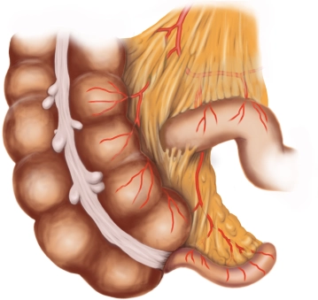
The large intestine (colon) is approximately 1.5 m long and begins with the junction of the small intestine into the cecum. The cecum is located at the level and below the ileocecal valve, has its own mesentery (→ mobility) containing the appendicular artery/vein (← ileocolic artery ← superior mesenteric artery), and is about 7 cm long. The blind-ending vermiform appendix is located on the dorsomedial wall of the cecum, just below the ileocecal valve in the taenia libera. It is intraperitoneal, and its length varies between 2-20 cm, with a diameter between 0.5 and 1 cm. Normally, the appendix runs from the posterior middle of the cecum to the middle of the body, but its position can be very variable, and thus also the location and intensity of tenderness. This surgical condition is mistakenly referred to as "appendicitis," although from an anatomical perspective, the affected organ is merely the vermiform appendix of the cecum.
Positional Variants of the Appendix:
- Descending type: Appendix extends into the small pelvis. In women, it can come into close proximity with the ovary.
- Medial position: Appendix lies between loops of the small intestine.
- Lateral position: Appendix lies between the lateral abdominal wall and the cecum.
- Retrocecal position: Appendix is folded upward behind the cecum (65%).
- Anterocecal position: Appendix is folded upward in front of the cecum.
- The subhepatic position: Appendix is folded upward towards the liver and is connected with it.
Histologically, the mucosa of the vermiform appendix shows the same structure as that of the large intestine. However, it contains a large number of lymphatic cells and thus becomes part of the immune system in humans. Additionally, in the wall of the appendix, the three separate strips of longitudinal muscle typical for the colon (taeniae) unite again into a complete layer.
Follow this link for more information on the anterior abdominal wall.