Appendectomy, open

Reading time readingtime 14:19 min.
  1. Skin incision

    Video
    Skin incision

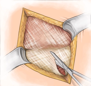
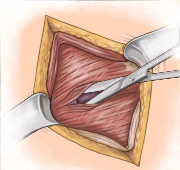
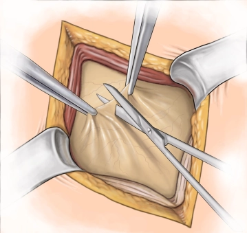
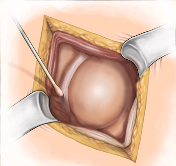
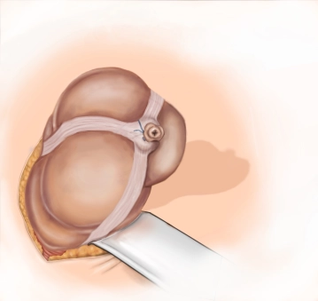
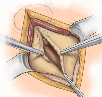
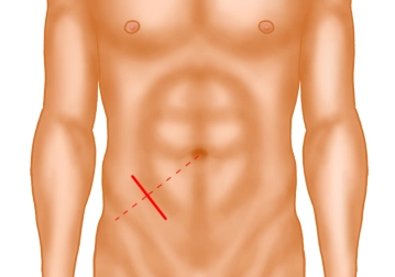
    A transverse incision approximately 3-4 cm long is made in the right lower abdomen (on an imaginary line between the anterior superior iliac spine and the navel in the caudal third). Subsequently, the subcutis and Scarpa's fascia are incised with the electric knife.

    Note: The transverse incision should be the standard approach, as the "curtain phenomenon" of the abdominal wall practically prevents incisional hernias, unlike the pararectal incision. The often-cited criticism that a transverse incision cannot be extended if the surgical approach changes is incorrect: The transverse incision can be easily extended cranially as a "hockey stick incision" or medially as a modified "Pfannenstiel incision".

  2. Splitting of the external aponeurosis

    Video
    Splitting of the external aponeurosis

    The external aponeurosis is incised with scissors from laterocranial to mediocaudal.

  3. Splitting of the muscle

    Video
    Splitting of the muscle

    The internal oblique and transversus muscles are bluntly split with scissors and retracted with Roux hooks.

  4. Peritoneal incision

    Video
    Peritoneal incision

    The peritoneum is exposed and incised with scissors.

  5. Swab collection

    Video
    Swab collection

    An intraperitoneal swab is taken.

  6. Luxation of the cecal pole

    Video
    Luxation of the cecal pole

    The cecal pole, along with the appendix, is grasped with a moist gauze and gently luxated in front of the abdominal wall with minimal traction.

  7. Skeletonization of the appendix

    Video
    Skeletonization of the appendix

    The skeletonization of the appendix is performed with Overholt clamps (near the appendix, Overholt 19 or 14) with stepwise transection and ligation (Vicryl 3-0) of the vessels of the mesoappendix.

  8. Placement of the purse-string suture

    Video
    Placement of the purse-string suture

    The purse-string suture necessary for the later inversion of the appendix is placed approximately 10 mm from the base using Vicryl 3-0 SH.

    Tip: In cases of difficult local findings, it may be helpful to leave the purse-string suture thread slightly longer opposite the entry and exit points when placing it. This facilitates the inversion of the stump by lifting the cecal pole at both ends of the thread and this loop.

  9. Ligation and resection of the appendix base

    Video
    Ligation and resection of the appendix base

    The appendix is ligated at the base (Vicryl 3-0) and excised with a knife (discarded) while lying on the Braunol handle.

  10. Inversion of the stump

    Video
    Inversion of the stump

    The purse-string suture is tied, and the appendix stump is inverted with forceps (assistant) (discard forceps and scissors after cutting the thread).
    Subsequently, the Douglas pouch is dabbed first moist, then dry.

    Note: No routine drainage with a sanitized focus and local peritonitis!

    Remark: "Meckel" only with relatively bland appendix.

    • Meckel's diverticulum:
      Clamp the diverticulum sufficiently far from its base, excise, and suture transversely, single-layer extramucosal (monofilament 4-0) or excision with a stapling device.
  11. Peritoneal and fascial suture

    Video
    Peritoneal and fascial suture

    The peritoneum (2-0) and the fascia (0) are closed with monofilament, absorbable material.

  12. Ending the operation

    Video
    Ending the operation

    Subcutaneous suture, skin suture.