Anatomy - EVAR – Endovascular repair of abdominal aortic aneurysm (Y-graft) – Vascular Surgery

  1. Abdominal aorta

    Abdominal aorta
    Zum Vergrößern bitte klicken

    Origin

    • It is the extension of the thoracic aorta after it passes through the aortic hiatus at the level of 12th thoracic vertebra (T12)

    Course

    • Retroperitoneal
    • Left of midline, anterior to spine
    • At the level of umbilicus/4th lumbar vertebra (L4) dividing (aortic bifurcation) into the common iliac arteries

    Cephalocaudal branches

    • Inferior phrenic arteries
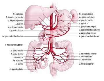
    • Celiac trunk
    • Middle suprarenal artery (left and right)
    • Superior mesenteric artery
    • Renal artery (left and right)
    • Ovarian / testicular artery (left and right)
    • Lumbar arteries
    • Inferior mesenteric artery
    • Median sacral artery

    Distribution

    • Paired branches: abdominal wall, paired retroperitoneal organs (kidneys, adrenal glands), gonads
    • Unpaired branches: spleen, liver, pancreas, unpaired digestive organs
  2. Paired branches of the abdominal aorta

    Main branches

    Course

    Branches and their distribution

    Inferior phrenic artery

    o    Courses craniad on inferior aspect of diaphragm toward its dome

    o    Reticular division into several terminal branches

    Superior suprarenal artery: adrenal gland

    Terminal branches: Inferior aspect of diaphragm

    Middle suprarenal artery

    o    Courses laterad to the adrenal glands

    Terminal branches: adrenal glands
    Renal artery

    o    Arises at around the level of the first / second lumbar vertebra (L1-2)

    o    Right renal artery: courses posterior to inferior vena cava and head of pancreas to right kidney

    o    Left renal artery: courses posterior to body of pancreas to left kidney

    Inferior suprarenal artery: adrenal gland

    Ovarian artery, 

    Testicular artery

    o    Arises inferior to the renal arteries /second lumbar vertebra (L2) from anterior aspect of aorta

    o    Crosses over psoas muscle, ureter and external iliac arteries to suspensory ligament of ovary / deep inguinal ring and testes

    Ovarian artery ovary, parts of uterine tube

    Testicular artery: testes, epididymides

    Lumbar arteries (bilateral 4)

    o    Run laterad posterior to psoas muscle over 1st to 4th lumbar vertebra (L1–4)

    o    Anastomose with arteries of the anterior abdominal wall (superior and inferior epigastric arteries, iliolumbar artery, deep circumflex iliac artery)

    Spinal branches: spinal canal

    Terminal branches: abdominal wall, muscles of the back


  3. Unpaired branches of the abdominal aorta

    • All unpaired branches of the abdominal aorta form anastomoses maintaining organ perfusion via collaterals:
      • Celiac trunk→ gastroduodenal artery→ superior mesenteric artery
      • Superior mesenteric artery -> middle and left celiac artery -> inferior mesenteric artery (“Riolan anastomosis”, marginal artery of Drummond, rarely accessory intermesenteric anastomosis (Williams and Klop)))
      • Inferior mesenteric artery→ superior and middle rectal artery→ internal iliac artery
    1. Celiac trunk
    • Arises at the level of the 12th thoracic vertebra (T12)
    • Divides into splenic artery, left gastric artery and common hepatic artery (largest-caliber branch of trunk)


    Main branches

    Course

    Branches and their course 

    Distribution

    Splenic artery

    o    Along superior margin of pancreas

    o    Enters the splenorenal ligament and spleen

    Pancreatic branches:

    o    Posterior to pancreas

    Dorsal pancreatic artery:

    o    Posterior to pancreas

    o    Anastomoses with greater pancreatic artery and continues as inferior pancreatic artery

    Greater pancreatic artery:

    o    Caudad along the posterior aspect of tail of pancreas

    o    Anastomosis at pancreatic tail with dorsal pancreatic artery and inferior pancreatic artery

    Pancreas
      

    Posterior gastric artery:

    o    Runs craniad in phrenicosplenic ligament to gastric fundus

    Stomach (posterior aspect of fundus)
      

    Short gastric arteries:

    o    Course in gastrosplenic ligament to gastric fundus

    Stomach (fundus and greater curvature)
      

    Left gastro-omental artery:

    o    Courses in gastrocolic ligament to greater curvature

    o    Anastomosis with right gastro-omental artery

    Stomach (fundus and greater curvature)
    Left gastric artery

    o    Runs to lesser curvature of stomach

    o    Anastomosis with right gastric artery

    Direct branchesStomach (lesser curvature)
      Esophageal branchesEsophagus
    Common hepatic artery

    o    Runs right to hepatic portal

    o    Divides into proper hepatic artery and gastroduodenal artery

    Proper hepatic artery

    o    Main branch of common hepatic artery

    o    Arises posterior to pylorus

    o    Runs in hepatoduodenal ligament together with hepatic portal vein and common bile duct to hepatic portal

    o    Gives rise to three branches:


    1. Right gastric artery:

    o    Runs in lesser omentum to lesser curvature of stomach

    o    Anastomosis with left gastric artery

    Stomach (right lesser curvature)
      2. Right hepatic artery

    Liver (right half)

    Gallbladder (cystic artery)

    Part of caudate lobe

      3. Left hepatic artery

    Liver (left half)

    Part of caudate lobe


    Gastroduodenal artery

    o    Courses posterior to lesser omentum to pancreas

    o    Divides into 2 branches:


    1. Right gastro-omental artery:

    o    Runs to greater curvature

    o    Anastomosis with left gastro-omental artery

    Pancreas, duodenum, stomach
      

    2. Anterior and posterior superior pancreaticoduodenal arteries:

    o    Form a vascular plexus around the pancreatic head with branches of the inferior pancreaticoduodenal artery

     
    2. Superior mesenteric artery
    • Arises from aorta at the level of L1-2
    • Courses anteroinferiorly
    • Branches: Branches: inferior pancreaticoduodenal artery, jejunal arteries and iliac arteries, ileocolic artery, right colic artery, middle colic artery

    Main branches

    Course

    Branches

    Distribution

    Inferior pancreaticoduodenal artery

    o    At horizontal part of duodenum it runs craniad to pancreatic head

    Anterior and posterior branch:

    o    Anastomosis with anterior and posterior superior pancreaticoduodenal artery respectively

    Pancreatic head
    Jenunal and iliac arteries

    o    Course in the mesenteric root to jejunum and Ileum

    o    Form a vascular arch

    Terminal branches (Aa. rectae)Jejunum, Ileum
    Ileocolic artery

    o    Runs in mesentery to ileocecal region

    Anterior and posterior cecal arteryCecum
      Appendicular artery (variant: branch of the posterior cecal artery)Vermiform appendix
    Right colic artery

    o    Runs in ascending mesocolon to ascending colon

    Divides into 2 branches that anastomose with the middle colic artery and ileocolic artery respectivelyAscending colon
    Middle colic artery

    o    Runs in transverse mesocolon to transverse colon

    Divides into 2 branches: right branch anastomoses with right colic artery (right flexure), left branch with left colic artery (left flexure)Transverse colon
    3. Inferior mesenteric artery
    • Arises from the aorta at the level of L3
    • Supplies the colon beyond the left flexure with left colic artery, sigmoid arteries and superior rectal artery

    Main branches

    Course

    Branches

    Distribution

    Left colic artery

    o    Runs in descending mesocolon to descending colon

    Divides into 2 branches that anastomose with the middle colic artery and sigmoid arteries respectivelyDescending colon
    Sigmoid arteries

    o    2-4 arteries forming a vascular arch in the sigmoid mesocolon

    o    Anastomosis with left colic artery and superior rectal artery

    Several small branches to sigmoid colonSigmoid colon
    Superior rectal artery

    o    Joins the rectum from posterior

    o    Anastomosis with middle rectal artery

    Several small branches to rectumUpper rectum
Arterial pelvic blood supply

The abdominal aorta divides at the aortic bifurcation (around level L4) into both common iliac arte

Activate now and continue learning straight away.

Single Access

Activation of this course for 3 days.

€4.99 inclusive VAT

Most popular offer

webop - Savings Flex

Combine our learning modules flexibly and save up to 50%.

from €3.29 / module

€39.50 / yearly payment

price overview

vascular surgery

Unlock all courses in this module.

€7.42 / month

€89.00 / yearly payment