Start your free 3-day trial — no credit card required, full access included

Perioperative management - EVAR – Endovascular repair of abdominal aortic aneurysm (Y-graft) – Vascular Surgery

  1. Indications

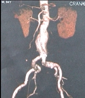
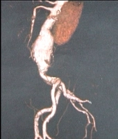
    • Abdominal aortic aneurysm (AAA) > 5 cm
    • Smaller aneurysms with eccentric AAA or ulceration with contained perforation (PAU, primary aortic ulcer)
    • Symptomatic aneurysms of any size

    Special indications and treatment options for Behçet disease, Marfan syndrome, mycotic aneurysms, such as, e.g., salmonella infections, and AAA with peripheral embolization.

    The indication for open repair of abdominal aortic aneurysm (AAA) basically results from comparing the patient's individual risk of rupture in the spontaneous course of the disease with the risk of open surgery. If the risk of spontaeous rupture over the course exceeds the individual surgical risk, open surgery is usually indicated.

    Rupture risk classification 
    FactorsLow riskModerate riskHigh risk
    Aneurysm diameter<5 cm5–6 cm>6 cm
    Growth rate per year<0,3 cm0,3–0,5 cm>0,5 cm
    Smoking/COPDLowModerateHigh
    Family historyNoneIsolatedCommon
    Arterial hypertensionNoneWell controlledUnstable despite treatment
    MorphologyFusiformSaccularEvaginations
    Gender MaleFemale
    Indication for surgery
    ClassificationSizeWallPresentationIndication for surgery
    Asymptomatic infrarenal

    >5 cm ♂

    >4.5 cm ♀

    IntactNoneElective
    Asymptomatic supraaortic >6 cmIntactNoneElective
    SymptomaticIndependently of other factorsIntactSpontaneous pain; tenderness in abdomen, back or sideUrgent, within 24 hours
    RupturedIndependently of other factorsContained or free ruptureDiffuse severe spontaneous pain / tenderness of tense abdomen, with/without hemorrhagic shockEmergency
      Aortoduodenal fistulaIntermitted vomiting, melenaEmergency
      Aortocaval fistulaRight heart failure, fistula bruit, truncal cyanosis, concurrent contrast enhancement of aorta & inferior vena cavaEmergency

    There are two types of repair in AAA:

    • open replacement of the abdominal aorta with a straight (tubular) or bifurcation graft (OAR, open aortic repair)
    • endovascular placement of a stent graft (EVAR, endovascular aortic repair)

    Laparoscopic aneurysm repair surgery, usually performed in combination with mini-laparotomy, is of lesser importance.

    The following recommendations serve as a guide to choosing between OAR and EVAR:

    OAR (trans-/retroperitoneal)

    • Normal life expectancy
    • Low surgical risk (fitness)
    • Anatomy unsuitable for EVAR: landing zone; aneurysm neck (angle, length); iliac vessels (stenosis, elongation, kinking); thrombi; calcification
    • Marfan and other connective tissue disorders

    EVAR (standard /customized graft)

    • Prior abdominal surgery
    • Limited life expectancy
    • High surgical risk
    • Anatomy suitable for EVAR (see above)

    Since stent graft systems are often of large caliber, EVAR requires adequate iliac artery lumen for access. Iliac arteries with atherosclerotic stenosis, tortuosity, kinking, or aneurysmal dilation are a problem.

    In the long run, endovascular aortic grafts are associated with a higher complication rate than open aortic surgery.

    The BAR Score Calculator→ www.britishaneurysmrepairscore.com can quickly calculate patient mortality risk for EVAR or OAR, which can be useful when advising patients about the risk of elective EVAR or OAR.

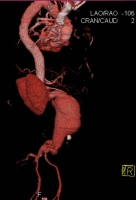
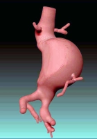
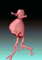
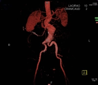
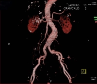
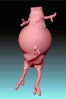
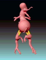
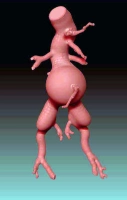
    The video example is an infrarenal, contained AAA with a diameter of > 5 cm:

    PM 312-1
    Figure 1: Preoperative anteroposterior angio-CT


    PM 312-2
    Figure 2: Preoperative lateral angio-CT


  2. Contraindications

    Since interventional procedures may require conversion to open surgery due to unexpected technical problems, preoperative risk assessment and definition of contraindications are important:

    • Heart failure (ejection fraction < 25-30%)
    • COPD (FEV1 < 0.8)
    • Refractory coronary heart disease
    • Incurable cancer

    Relative Contraindication:

    • Compensated renal failure (due to intraoperative contrast agent exposure)

    If the operation is to proceed in borderline cases despite contraindications, this must be discussed in detail with the patients and their relatives.    

    Technical contraindications to endovascular repair: see Preoperative diagnostic work-up/planning.

  3. Preoperative diagnostic work-up

    Thorough vascular surgery examination:

    • Pulse status
    • Doppler study of both legs
    • Possibly walk test in case of concomitant PAOD and poor foot pulses on palpation
    • Abdominal ultrasonography
    • Carotid color doppler study
    • Echocardiography
    • Exercise ECG
    • Laboratory panels (electroytes, coagulation, renal function, blood count, blood lipids)
    • Chest X-ray
    • Spiral CT
    • In case of concomitant PAOD or critical visceral artery morphology, possibly DSA of the abdominal aorta and pelvic and leg arteries

    Preoperative angiographic workup to clarify access pathology is especially important:

    • Stenosis of the femoral and pelvic arteries
    • Access artery aneurysms
    • Local infections such as, e.g., erythrasma
    • Previous operations in the surgical field

    The presence of a horseshoe kidney in AAA is a special case requiring special planning in both planned open surgery and endoluminal procedures. This planning must take into account the preservation of the multiple renal arteries in the vicinity of the abdominal aortic aneurysm.

    Below is a summary of the parameters to be addressed in the preoperative diagnostic work-up:

    1. Aortic pathology

    2. Risk factors (multimorbidity)

    3. Acess pathology 

    Planning

    Here, it is of crucial importance to obtain the exact dimensions of the aneurysm and take account of any calcification and stenosis in order to verify local suitability.

    PM 312-3
    Fig. 3: Preoperative AAA measurements

    One possible contraindication to endoluminal repair is severe kinking of the aneurysm neck. Moreover, coiling, compression and elongation have to be taken into account because the graft - depending on the centerline - may differ from the true aneurysm configuration and therefore be malaligned

    After analyzing the morphology, it should also be decided via which side to introduce the main body of the graft or whether to primarily aim for a cross-leg ballerina configuration in order to facilitate probing of the contralateral leg.  

    PM 312-4
    Fig. 4: Cross-leg ballerina configuration

     Examples of particular aneurysm configurations and access pathologies

    PM 312-5
    Figure 5: Kinking of the aneurysm neck


    PM 312-6
    Figure 6: Left convex AAA


    PM 312-7
    Figure 7: Right convex AAA


    PM 312-8
    Figure 8: AAA elongations


    PM 312-9
    Figure 9: Dilated common iliac artery


    PM 312-10
    Figure 10: Pararenal AAA


    PM 312-11
    Figure 11: Stenosis of the iliac arteries


    PM 312-12
    Figure 12: Triple aneuyrsma
    PM 312-13
    Figure 13: Spherical AAA
  4. Special preparation

    • Identify blood group, provide for packed RBCs, if needed
    • Remove the hair in the surgical field
    • Possibly, place urinary catheter
    • Prophylactic antibiotics are usually administered as recommended by the German Society for Vascular Surgery (single-shot cefuroxime 30 minutes before skin incision), but the benefit is currently subject to debate because of issues around antibiotic resistance (Robert-Koch-Institute).   
  5. Informed consent

    General surgical risks

    • Major bleeding, blood transfusions, transmission of hepatitis/HIV through allogeneic blood transfusions
    • Allergy/intolerance
    • Wound infection
    • Thrombosis/embolism
    • Skin/vascular/nerve damage, e.g., due to patient positioning
    • Keloids (in open surgery)

    Specific procedural risks

    • Vascular injuries, e.g., during graft delivery: surgical hemostasis, blood transfusions, in case of massive bleeding or aortic rupture, immediate conversion to open surgery
    • Pseudoaneurysm of the punctured vessels, arteriovenous fistula, seroma
    • Primary aortoenteric fistula: surgical intervention, possibly with removal of the vascular graft
    • Inadequate graft fixation or leakage: corrective procedures, possibly open surgery
    • Graft infection: possible after days, months, or even years; endocarditis, sepsis, leg ischemia, amputation; surgical removal of vascular graft
    • Injury of adjacent organs; intestinal ischemia → resection, stoma
    • Nerve lesions→ dysesthesia; pain; paralysis of abdominal wall / thigh muscles
    • Lymph fistula
    • Temporary or permanent lymphedema of the legs; compression stockings, lymphatic drainage
    • Secondary bleeding
    • Impotence    
    • Deterioration of renal function induced by intraoperative angiography, chronic dialysis

    Risks due to impaired perfusion

    • Thrombosis/embolism: pulmonary embolism, apoplexy, myocardial infarction; prophylaxis: heparin → HIT II risk
    • Legs: thrombosis of the graft and possibly adjacent vascular segments, possibly leg ischemia, amputation (e.g., toes)
    • Kidneys: due to thrombosis or graft delivery; despite immediate surgical repair, kidney damage may be permanent → dialysis
    • Bowel: impaired perfusion due to thrombosis/embolism and possibly graft delivery; vascular surgery required, possibly bowel resection, chronic stoma
    • Spinal cord: depending on the size of the aneurysm, impaired perfusion due to graft delivery; temporary paresthesia/paresis, possibly also permanent paraplegia if the artery of Adamkiewicz originates low in the aorta
    • Gluteal muscles: due to overstenting of the iliac arteries, in particular bilaterally; claudication during walking, possibly gluteal necrosis
    • Liver: Impaired function due to hypoperfusion, especially in pre-existing liver disease.
  6. Anesthesia

  7. Positioning

    PM 312-14

    Supine, with both arms abducted. The upper body should be slightly retroflexed at the level of the thoracoabdominal transition between the pubic bone and xyphoid. Protect both feet with cotton padded shoes (caution: pressure injury).

  8. Operating room setup

    PM 312-15

    Depending on the configuration of the aneurysm and from which side the main body of the endograft is inserted, the surgeon stands alternately on the right and left, with the assistant opposite in each case. The scrub nurser stands next to the surgeon, thus also changing position as required

  9. Special instruments and fixation systems

    • Smal vascular instrument tray
    • Adequate guide wires (Terumo®, Lunderquist®, Amplatz®)
    • Guiding catheters (e.g. Hockey Stick®, Renal Double Curve®, Sidewinder®, etc.)
    • Angioplasty balloons in different sizes (6-9 mm)
    • Stents for renal arteries and possibly for additional graft fixation
    • Long and short pigtail catheters, pigtail with gradations
    • Super compliant balloon catheter with at least 20 ml capacity
    • DSA unit, high pressure injector for contrast media
    • Suitable endoluminal graft customized to preoperative measurements
    • Have instruments ready for conversion to emergent open surgery
  10. Postoperative management

    Postoperative analgesia:

    Follow this link to PROSPECT (Procedures Specific Postoperative Pain Management) and to the current German guideline Behandlung akuter perioperativer and posttraumatischer Schmerzen [Treatment of acute perioperative and posttraumatic pain].

    Postoperative care

    • 24-hour monitoring on ICU or possibly intermediate care unit
    • Regular ward from postoperative day 1 if possible
    • Close cardiovascular and pulmonary monitoring
    • Coagulation monitoring (e.g., for DIC)
    • Check pulse in legs and capillary perfusion in feet

    Deep venous thrombosis prophylaxis

    • Weight-adjusted low-molecular weight heparin; in multifocal arteriosclerosis ASA 100 mg/day when oral nutrition is restarted
    • for patients on phenprocoumon, switch from heparin to phenprocoumon on postoperative day 5

     Ambulation

    • To edge of bed from postoperative day 1-2

    Physical therapy

    • Isometric and breathing exercises    

    Diet

    • 4– 6 hours after surgery

    Bowel movement

    • On day 3 enema if no spontaneous bowel movement

    Work disability

    • Around 3 months