Start your free 3-day trial — no credit card required, full access included

Perioperative management - Percutaneous transluminal angioplasty (PTA) and stenting in bilateral renal artery stenosis - Vascular surgery

  1. Indications

    PM 316-1
    • High grade unilateral or bilateral RAS (>70%) with the aim of organ preservation (see indication in video clip)
    • RAS in single kidney function
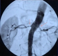
    • Fibromuscular dysplasia of the renal arteries with stenosis and hypertension
    • Difficult to control arterial hypertension in RAS

    Angioplasty is not indicated in asymptomatic renal artery stenosis (even if bilateral).

    In unilateral stenosis and impaired global renal function, the kidney downstream of the stenosis has usually been spared the elevated blood pressure, whereas the kidney with normal arterial lumen has been impaired by chronic hypertension (renin–angiotensin mechanism). Often, renal artery angioplasty of the unilateral stenosis can then improve the function of the poststenotic kidney. 

    Revascularization of the renal artery is not promising in kidneys with a longitudinal diameter of less than 8 cm and/or proteinuria >1 g/day.

    (See also the German guideline Leitlinie - S2K Erkrankungen der Nierenarterie) [Consensus S2 Guideline - Disorders of the renal artery)

  2. Contraindications

    • ASA IV
    • Incurable malignancy
    • Endovascular intervention: unsuitable access vessels
  3. Preoperative diagnostic work-up

    Detailed angiography for vascular surgery:

    • Pulse status, Doppler status of legs, color flow Doppler ultrasonography of carotid arteries
    • Abdominal duplex ultrasonography with blood flow study in renal artery and parenchyma
    • Duplex ultrasonography of potential access vessels
    • Separate renal clearance for each kidney (scintigraphy)
    • MR angiography of renal aortic segment (direction of the renal arteries → cephalic, caudal)

    Also:

    • Chest X-ray
    • Echocardiography (left ventricular function and mural thickness)
    • ECG, possibly exercise ECG (signs of ischemia)
    • Laboratory panels (RBC; coagulation; blood lipids; electrolytes; renal function test)
    • Clinical examination of the access region (e.g. inguinal infection)
  4. Special preparation

    • Mark side of RAS on patient before surgery
    • Depilation of abdomen and both groins
    • Have patient fast for 6 hours preoperatively
    • Since procedure may last several hours → Foley catheter (if procedure is under local anesthetic, a painfully full urinary bladder makes the patient restless, and can also trigger vagal and cardiac sensations)
  5. Informed consent

    • Change of access route, e.g. transfemoral -> transcubital
    • Allogeneic blood transfusion, hepatitis, HIV
    • Allergic reaction, e.g. contrast media
    • Contrast-agent induced nephropathy, possibly lifelong dialysis
    • Hematoma/ secondary bleeding from groin puncture site
    • Vascular dissection in the femoral or iliac arteries, possibly requiring open surgical repair
    • AV fistula formation, pseudoaneurysm in the femoral artery requiring surgical repair
    • Inguinal infection, deep infection, sepsis
    • Injury to the femoral nerve
    • Failed renal artery dilation with perforation or dissection → conversion of surgical aproach to open abdominal exposure, vein graft harvesting, e.g., leg
    • Perforation of renal parenchyma and renal hemorrhage requiring open exposure; possibly nephrectomy, if hemorrhage cannot be controlled
    • Stent malalignment, secondary stent migration→ possibly surgical repair
    • Elimination of RAS → hypotension, cerebral ischemia (rare)  
  6. Anesthesia

  7. Positioning

    PM 316-2
    • Supine, with arm abducted
  8. Operating room setup

    PM 316-3

    The surgeon stands on the side to be operated on (= in video clip puncture of the left femoral artery), with the assistant facing him/her. The scrub nurse stands to the right of the assistant. The C-arm for intraoperative angiography approaches from the right side, with the monitor positioned to the right of the patient's head and clearly visible to the surgeon.

  9. Special instruments and fixation systems

    • Mobile or stationary DSA unit or hybrid operating room
    • High pressure contrast media injector
    • Various sheaths(6F and larger)
    • Standard instrument set for vascular surgery, retractor
    • Various guidewires (Terumo®, soft-tipped stiffer wires specifically for renal arteries)
    • Various guiding catheters (such as RDC = Renal Double Curve, Hockey Stick, SIM 1 and 2 = Simmons type sidewinder)
    • Dilation balloons in different sizes (3–6 mm)
    • Balloon-expandable nitinol stents (4–6 mm, length 20 mm, e.g. Palmaz Genesis®; nitinol = nickel-titanium alloy) for renal angioplasty
    • Have instruments ready for conversion to emergent open surgery

    Caution:

    Renal artery interventions require longer wires and catheters than interventions in the iliac arteries or abdominal aorta and special guiding catheters (e.g., RDC, Hockey Stick, SIMS 1 and 2).

  10. Postoperative management

    Postoperative analgesia:

    Follow this link to PROSPECT (Procedures Specific Postoperative Pain Management) and to the current German guideline Behandlung akuter perioperativer and posttraumatischer Schmerzen [Treatment of acute perioperative and posttraumatic pain]

    Postoperative care

    • Weight-adapted intravenous heparin (at least 10,000 IU/24 hours) for 3 days overlapping with platelet aggregation inhibitors
    • Long-term treatment with ASA 100 mg/d, plus clopidrogel 75 mg/d for 4 weeks
    • If patient is being treated for CHD or arrhythmias order cardiology consult
    • 24-hour monitoring: continuous ECG, peripheral O2 level, NIBP monitoring (->risk of  excessive drop in blood pressure)
    • Check renal function parameters
    • Routine follow-up study with duplex ultrasonography

    Ambulation

    • After 24 hours

    Physical therapy

    • Not required

    Diet

    • After 4 hours

    Bowel movement

    • Not required

    Work disability

    • Around 1 week