Start your free 3-day trial — no credit card required, full access included

Anatomy - Percutaneous transluminal angioplasty (PTA) and stenting in bilateral renal artery stenosis - Vascular surgery

  1. Abdominal aorta

    Abdominal aorta
    Zum Vergrößern bitte klicken

    Origin

    • It is the extension of the thoracic aorta after it passes through the aortic hiatus at the level of 12th thoracic vertebra (T12)

    Course

    • Retroperitoneal
    • Left of midline, anterior to spine
    • At the level of umbilicus/4th lumbar vertebra (L4) dividing (aortic bifurcation) into the common iliac arteries

    Cephalocaudal branches

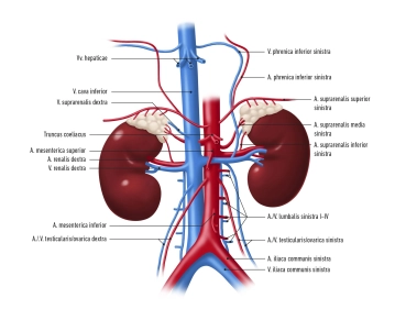
    • Inferior phrenic arteries
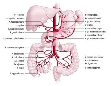
    • Celiac trunk
    • Left and right middle suprarenal artery
    • Superior mesenteric artery
    • Left and right renal artery
    • Left and right ovarian / testicular artery
    • Lumbar arteries
    • Inferior mesenteric artery
    • Median sacral artery

    Arterial blood supply for

    • Paired branches: abdominal wall, paired retroperitoneal organs, gonads
    • Unpaired branches: spleen, unpaired digestive organs
  2. Renal vasculature

    Renal vasculature
    Zum Vergrößern bitte klicken

    Arterial supply to the kidney is via the renal arteries originating directly from the aorta. Venous return is via the homonymous renal veins terminating in the inferior vena cava.

    After arising from the abdominal aorta the right renal artery crosses posterior to the inferior vena cava, while the left renal artery travels directly to the renal hilum. The reverse is true for the veins: The right renal vein travels directly to the inferior vena cava, whereas the left renal vein crosses anterior to the aorta.

    The renal artery divides shortly before the renal hilum into its principal branches. Around 18% of the population have more than one renal artery, 2–3% have even more than three renal arteries.

  3. Renal artery stenosis

    Narrowing of the arterial lumen by more than 70% is classified as hemodynamically significant renal artery stenosis (RAS). Underlying arteriosclerosis is implicated in around 90% of RAS cases, manifesting in particular at the aortic orifice and in the proximal third of the renal artery. This type is observed especially in older patients with cardiovascular risk factors (in particular: diabetes; hypertension; coronary heart disease; arterial occlusive disease) and is usually a progressive disorder.

    The second most common cause of RAS is fibromuscular dysplasia (FMD) seen in the younger population, in particular in premenopausal women aged 15–50 years, presenting in the distal two-thirds of the renal arteries. The prevalence of RAS in the German population is not known.

    The symptoms and sequelae of RAS may include ischemic nephropathy and renovascular hypertension. The latter cause is implicated in around 1–3% of hypertensive patients. How often renovascular disease is present in chronic renal failure is not known either. The incidence is estimated to be around 10–40%.